
Number of data: 7660
Dataset: all data
| pdb_000038fv | CYP3A4 bound to ritonavir | ||
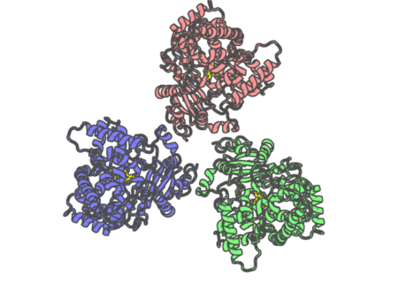 |
Experimental method | ELECTRON MICROSCOPY |
|---|---|---|
| Structural data | available | |
| Non-standard amino acids | None | |
| Missing residues/ Structural gap | None / None | |
| Resolution | None | |
| Deposition date | 2026-08-24 |
| pdb_000038fw | CYP3A4 bound to ketoconazole | ||
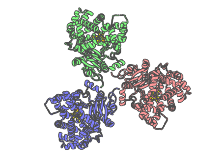 |
Experimental method | ELECTRON MICROSCOPY |
|---|---|---|
| Structural data | available | |
| Non-standard amino acids | None | |
| Missing residues/ Structural gap | Exists / Exists | |
| Resolution | None | |
| Deposition date | 2026-08-24 |
| pdb_000038fx | CYP3A4 bound to azamulin | ||
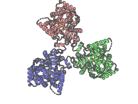 |
Experimental method | ELECTRON MICROSCOPY |
|---|---|---|
| Structural data | available | |
| Non-standard amino acids | None | |
| Missing residues/ Structural gap | Exists / Exists | |
| Resolution | None | |
| Deposition date | 2026-08-24 |
| pdb_000038fy | CYP3A4 bound to vardenafil | ||
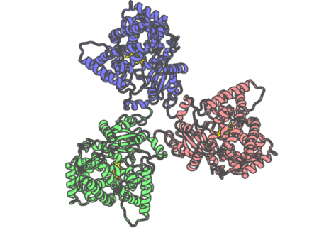 |
Experimental method | ELECTRON MICROSCOPY |
|---|---|---|
| Structural data | available | |
| Non-standard amino acids | None | |
| Missing residues/ Structural gap | None / None | |
| Resolution | 3.5 Å | |
| Deposition date | 2026-08-24 |
| pdb_00003a0b | Crystal structure of Br-substituted Photosystem II complex | ||
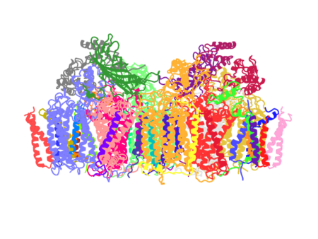 |
Experimental method | X-RAY DIFFRACTION |
|---|---|---|
| Structural data | available | |
| Non-standard amino acids | None | |
| Missing residues/ Structural gap | None / None | |
| Resolution | 3.7 Å | |
| Deposition date | 2009-03-16 |
| pdb_00003a0g | Crystal structure analysis of guinea pig oxyhemoglobin at 2.5 angstroms resolution | ||
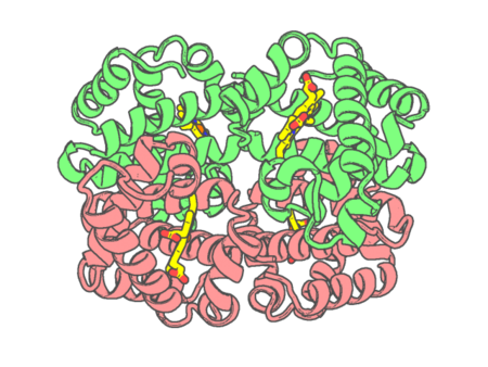 |
Experimental method | X-RAY DIFFRACTION |
|---|---|---|
| Structural data | available | |
| Non-standard amino acids | None | |
| Missing residues/ Structural gap | None / None | |
| Resolution | 2.5 Å | |
| Deposition date | 2009-03-17 |
| pdb_00003a0h | Crystal structure of I-substituted Photosystem II complex | ||
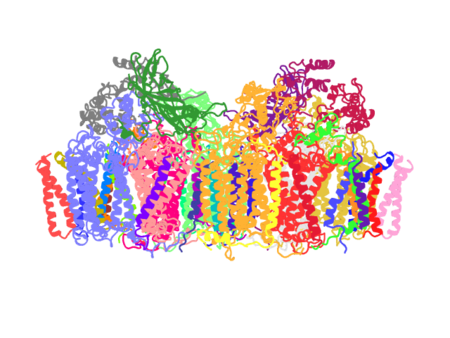 |
Experimental method | X-RAY DIFFRACTION |
|---|---|---|
| Structural data | available | |
| Non-standard amino acids | None | |
| Missing residues/ Structural gap | None / None | |
| Resolution | 4 Å | |
| Deposition date | 2009-03-17 |
| pdb_00003a15 | Crystal Structure of Substrate-Free Form of Aldoxime Dehydratase (OxdRE) | ||
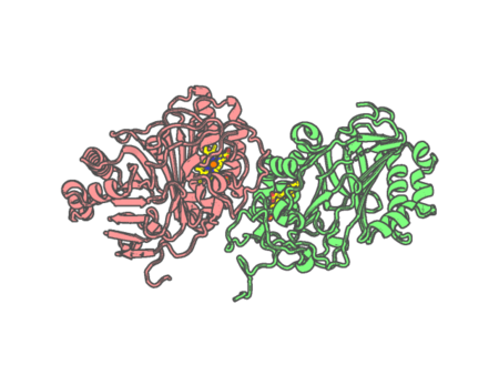 |
Experimental method | X-RAY DIFFRACTION |
|---|---|---|
| Structural data | available | |
| Non-standard amino acids | None | |
| Missing residues/ Structural gap | None / None | |
| Resolution | 1.79 Å | |
| Deposition date | 2009-03-26 |
| pdb_00003a16 | Crystal Structure of Aldoxime Dehydratase (OxdRE) in Complex with Propionaldoxime | ||
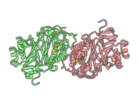 |
Experimental method | X-RAY DIFFRACTION |
|---|---|---|
| Structural data | available | |
| Non-standard amino acids | None | |
| Missing residues/ Structural gap | None / None | |
| Resolution | 1.6 Å | |
| Deposition date | 2009-03-26 |
| pdb_00003a17 | Crystal Structure of Aldoxime Dehydratase (OxdRE) in Complex with Butyraldoxime (Co-crystal) | ||
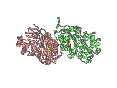 |
Experimental method | X-RAY DIFFRACTION |
|---|---|---|
| Structural data | available | |
| Non-standard amino acids | None | |
| Missing residues/ Structural gap | None / None | |
| Resolution | 2.5 Å | |
| Deposition date | 2009-03-26 |
| pdb_00003a18 | Crystal Structure of Aldoxime Dehydratase (OxdRE) in Complex with Butyraldoxime (soaked crystal) | ||
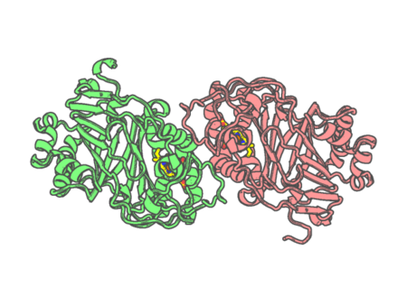 |
Experimental method | X-RAY DIFFRACTION |
|---|---|---|
| Structural data | available | |
| Non-standard amino acids | None | |
| Missing residues/ Structural gap | None / None | |
| Resolution | 1.8 Å | |
| Deposition date | 2009-03-26 |
| pdb_00003a1l | Crystal Structure of 11,11'-Dichlorochromopyrrolic Acid Bound Cytochrome P450 StaP (CYP245A1) | ||
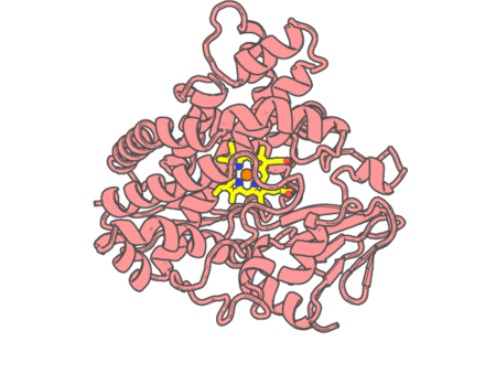 |
Experimental method | X-RAY DIFFRACTION |
|---|---|---|
| Structural data | available | |
| Non-standard amino acids | None | |
| Missing residues/ Structural gap | None / None | |
| Resolution | 2.5 Å | |
| Deposition date | 2009-04-09 |
| pdb_00003a2g | Crystal Structure of K102C-Myoglobin conjugated with Fluorescein | ||
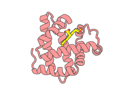 |
Experimental method | X-RAY DIFFRACTION |
|---|---|---|
| Structural data | available | |
| Non-standard amino acids | None | |
| Missing residues/ Structural gap | None / None | |
| Resolution | 1.75 Å | |
| Deposition date | 2009-05-20 |
| pdb_00003a4g | Structure of cytochrome P450 vdh from Pseudonocardia autotrophica (trigonal crystal form) | ||
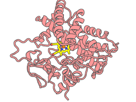 |
Experimental method | X-RAY DIFFRACTION |
|---|---|---|
| Structural data | available | |
| Non-standard amino acids | None | |
| Missing residues/ Structural gap | None / None | |
| Resolution | 1.75 Å | |
| Deposition date | 2009-07-07 |
| pdb_00003a4h | Structure of cytochrome P450 vdh from Pseudonocardia autotrophica (orthorhombic crystal form) | ||
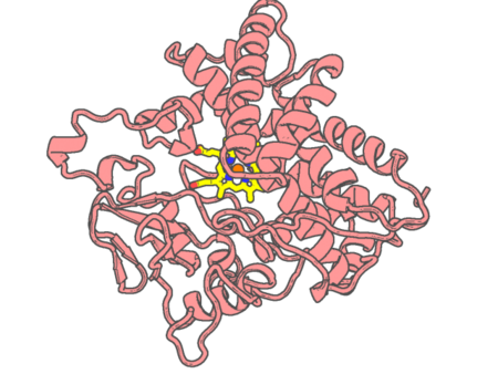 |
Experimental method | X-RAY DIFFRACTION |
|---|---|---|
| Structural data | available | |
| Non-standard amino acids | None | |
| Missing residues/ Structural gap | None / None | |
| Resolution | 3.06 Å | |
| Deposition date | 2009-07-07 |
| pdb_00003a4z | Structure of cytochrome P450 Vdh mutant (Vdh-K1) obtained by directed evolution | ||
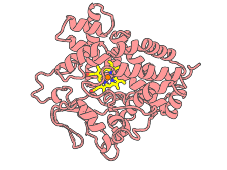 |
Experimental method | X-RAY DIFFRACTION |
|---|---|---|
| Structural data | available | |
| Non-standard amino acids | None | |
| Missing residues/ Structural gap | None / None | |
| Resolution | 2.2 Å | |
| Deposition date | 2009-07-24 |
| pdb_00003a50 | Structure of cytochrome P450 Vdh mutant (Vdh-K1) obtained by directed evolution with bound vitamin D3 | ||
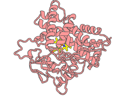 |
Experimental method | X-RAY DIFFRACTION |
|---|---|---|
| Structural data | available | |
| Non-standard amino acids | None | |
| Missing residues/ Structural gap | None / None | |
| Resolution | 2.05 Å | |
| Deposition date | 2009-07-24 |
| pdb_00003a51 | Structure of cytochrome P450 Vdh mutant (Vdh-K1) obtained by directed evolution with bound 25-hydroxyvitamin D3 | ||
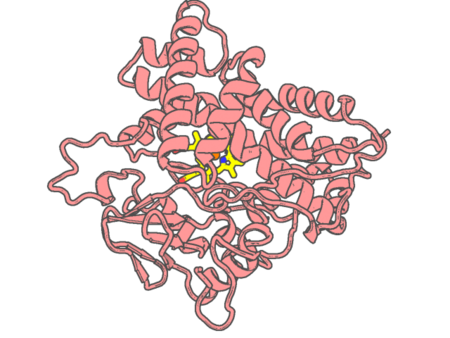 |
Experimental method | X-RAY DIFFRACTION |
|---|---|---|
| Structural data | available | |
| Non-standard amino acids | None | |
| Missing residues/ Structural gap | None / None | |
| Resolution | 2 Å | |
| Deposition date | 2009-07-24 |
| pdb_00003a59 | Structure of Hemoglobin from flightless bird (Struthio camelus) | ||
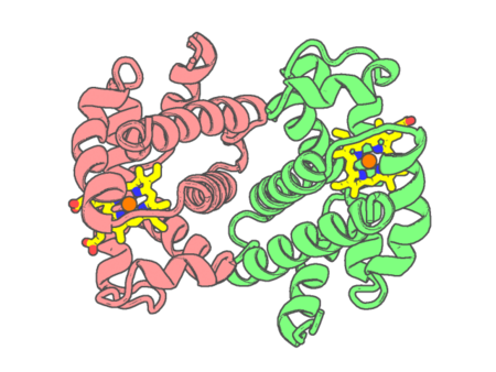 |
Experimental method | X-RAY DIFFRACTION |
|---|---|---|
| Structural data | available | |
| Non-standard amino acids | None | |
| Missing residues/ Structural gap | None / None | |
| Resolution | 3.41 Å | |
| Deposition date | 2009-08-03 |
| pdb_00003a5a | Crystal structure of a hemoglobin component V from Propsilocerus akamusi (pH5.6 coordinates) | ||
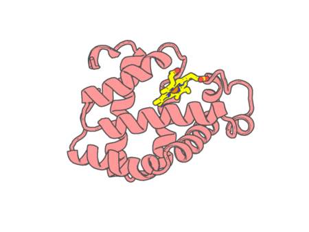 |
Experimental method | X-RAY DIFFRACTION |
|---|---|---|
| Structural data | available | |
| Non-standard amino acids | None | |
| Missing residues/ Structural gap | None / None | |
| Resolution | 1.83 Å | |
| Deposition date | 2009-08-05 |