
Number of data: 7660
Dataset: all data
| pdb_00003aeb | Crystal structure of porcine heart mitochondrial complex II bound with N-(3-Phenoxy-phenyl)-2-trifluoromethyl-benzamide | ||
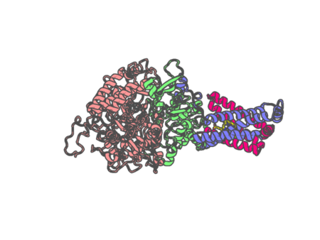 |
Experimental method | X-RAY DIFFRACTION |
|---|---|---|
| Structural data | available | |
| Non-standard amino acids | None | |
| Missing residues/ Structural gap | None / None | |
| Resolution | 3 Å | |
| Deposition date | 2010-02-04 |
| pdb_00003aec | Crystal structure of porcine heart mitochondrial complex II bound with 2-Iodo-N-(1-methylethyl)-benzamid | ||
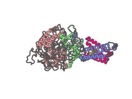 |
Experimental method | X-RAY DIFFRACTION |
|---|---|---|
| Structural data | available | |
| Non-standard amino acids | None | |
| Missing residues/ Structural gap | None / None | |
| Resolution | 3.61 Å | |
| Deposition date | 2010-02-04 |
| pdb_00003aed | Crystal structure of porcine heart mitochondrial complex II bound with 2-Iodo-N-phenyl-benzamide | ||
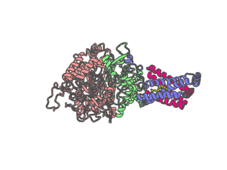 |
Experimental method | X-RAY DIFFRACTION |
|---|---|---|
| Structural data | available | |
| Non-standard amino acids | None | |
| Missing residues/ Structural gap | None / None | |
| Resolution | 3.52 Å | |
| Deposition date | 2010-02-04 |
| pdb_00003aee | Crystal structure of porcine heart mitochondrial complex II bound with Atpenin A5 | ||
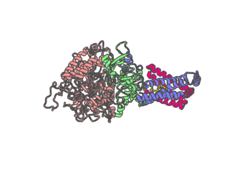 |
Experimental method | X-RAY DIFFRACTION |
|---|---|---|
| Structural data | available | |
| Non-standard amino acids | None | |
| Missing residues/ Structural gap | None / None | |
| Resolution | 3.22 Å | |
| Deposition date | 2010-02-04 |
| pdb_00003aef | Crystal structure of porcine heart mitochondrial complex II with an empty quinone-binding pocket | ||
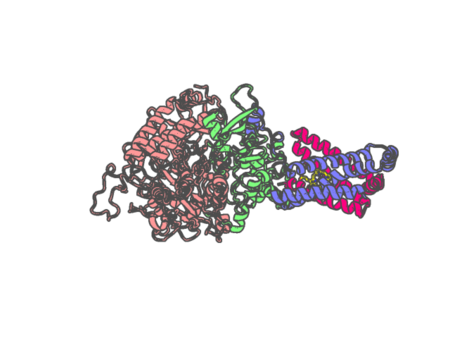 |
Experimental method | X-RAY DIFFRACTION |
|---|---|---|
| Structural data | available | |
| Non-standard amino acids | None | |
| Missing residues/ Structural gap | None / None | |
| Resolution | 2.8 Å | |
| Deposition date | 2010-02-04 |
| pdb_00003aeg | Crystal structure of porcine heart mitochondrial complex II bound with N-Biphenyl-3-yl-2-iodo-benzamide | ||
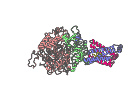 |
Experimental method | X-RAY DIFFRACTION |
|---|---|---|
| Structural data | available | |
| Non-standard amino acids | None | |
| Missing residues/ Structural gap | None / None | |
| Resolution | 3.27 Å | |
| Deposition date | 2010-02-04 |
| pdb_00003afv | Dye-decolorizing peroxidase (DyP) at 1.4 A resolution | ||
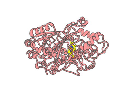 |
Experimental method | X-RAY DIFFRACTION |
|---|---|---|
| Structural data | available | |
| Non-standard amino acids | None | |
| Missing residues/ Structural gap | None / None | |
| Resolution | 1.4 Å | |
| Deposition date | 2010-03-11 |
| pdb_00003ag0 | Crystal structure of carbonmonoxy human cytoglobin | ||
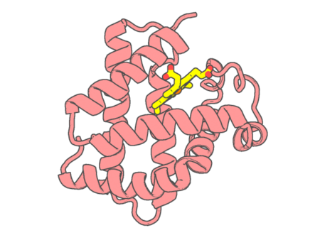 |
Experimental method | X-RAY DIFFRACTION |
|---|---|---|
| Structural data | available | |
| Non-standard amino acids | None | |
| Missing residues/ Structural gap | None / None | |
| Resolution | 2.6 Å | |
| Deposition date | 2010-03-17 |
| pdb_00003ag1 | Bovine Heart Cytochrome c Oxidase in the Carbon Monoxide-bound Fully Reduced State at 280 K | ||
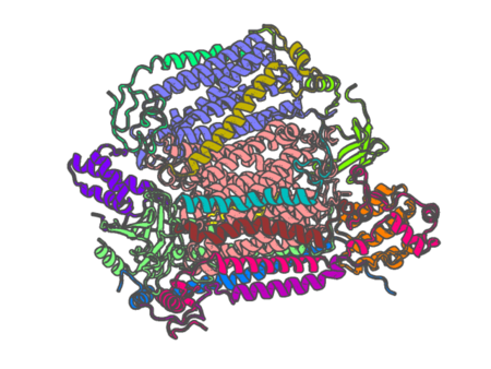 |
Experimental method | X-RAY DIFFRACTION |
|---|---|---|
| Structural data | available | |
| Non-standard amino acids | Included | |
| Missing residues/ Structural gap | None / None | |
| Resolution | 2.2 Å | |
| Deposition date | 2010-03-19 |
| pdb_00003ag2 | Bovine Heart Cytochrome c Oxidase in the Carbon Monoxide-bound Fully Reduced State at 100 K | ||
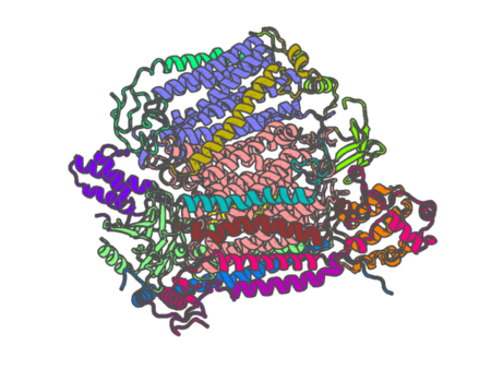 |
Experimental method | X-RAY DIFFRACTION |
|---|---|---|
| Structural data | available | |
| Non-standard amino acids | Included | |
| Missing residues/ Structural gap | None / None | |
| Resolution | 1.802 Å | |
| Deposition date | 2010-03-19 |
| pdb_00003ag3 | Bovine Heart Cytochrome c Oxidase in the Nitric Oxide-bound Fully Reduced State at 100 K | ||
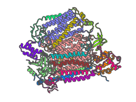 |
Experimental method | X-RAY DIFFRACTION |
|---|---|---|
| Structural data | available | |
| Non-standard amino acids | Included | |
| Missing residues/ Structural gap | None / None | |
| Resolution | 1.8 Å | |
| Deposition date | 2010-03-19 |
| pdb_00003ag4 | Bovine Heart Cytochrome c Oxidase in the Cyanide Ion-bound Fully Reduced State at 100 K | ||
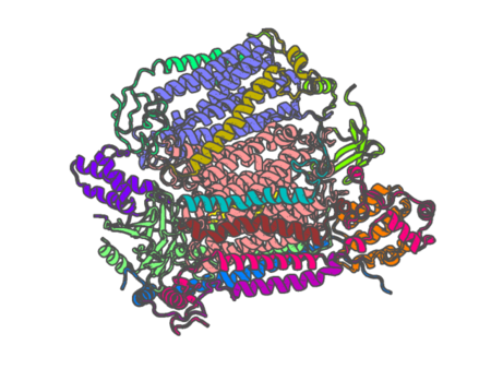 |
Experimental method | X-RAY DIFFRACTION |
|---|---|---|
| Structural data | available | |
| Non-standard amino acids | Included | |
| Missing residues/ Structural gap | None / None | |
| Resolution | 2.05 Å | |
| Deposition date | 2010-03-19 |
| pdb_00003aq5 | Crystal structure of truncated hemoglobin from Tetrahymena pyriformis, Fe(II)-O2 form | ||
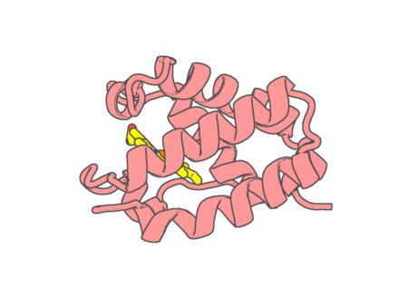 |
Experimental method | X-RAY DIFFRACTION |
|---|---|---|
| Structural data | available | |
| Non-standard amino acids | None | |
| Missing residues/ Structural gap | None / None | |
| Resolution | 1.78 Å | |
| Deposition date | 2010-10-25 |
| pdb_00003aq6 | Crystal structure of truncated hemoglobin from Tetrahymena pyriformis, Fe(III) form | ||
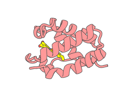 |
Experimental method | X-RAY DIFFRACTION |
|---|---|---|
| Structural data | available | |
| Non-standard amino acids | None | |
| Missing residues/ Structural gap | None / None | |
| Resolution | 1.93 Å | |
| Deposition date | 2010-10-25 |
| pdb_00003aq7 | Crystal structure of truncated hemoglobin from Tetrahymena pyriformis, Y25F mutant, Fe(III) form | ||
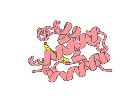 |
Experimental method | X-RAY DIFFRACTION |
|---|---|---|
| Structural data | available | |
| Non-standard amino acids | None | |
| Missing residues/ Structural gap | None / None | |
| Resolution | 1.77 Å | |
| Deposition date | 2010-10-25 |
| pdb_00003aq8 | Crystal structure of truncated hemoglobin from Tetrahymena pyriformis, Q46E mutant, Fe(III) form | ||
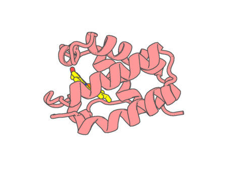 |
Experimental method | X-RAY DIFFRACTION |
|---|---|---|
| Structural data | available | |
| Non-standard amino acids | None | |
| Missing residues/ Structural gap | None / None | |
| Resolution | 1.83 Å | |
| Deposition date | 2010-10-25 |
| pdb_00003aq9 | Crystal structure of truncated hemoglobin from Tetrahymena pyriformis, Q50E mutant, Fe(III) form | ||
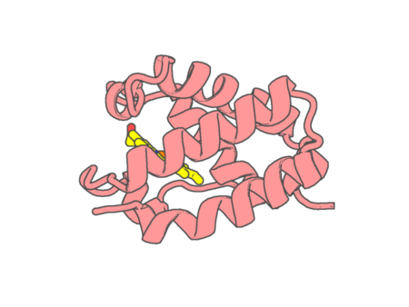 |
Experimental method | X-RAY DIFFRACTION |
|---|---|---|
| Structural data | available | |
| Non-standard amino acids | None | |
| Missing residues/ Structural gap | None / None | |
| Resolution | 1.74 Å | |
| Deposition date | 2010-10-25 |
| pdb_00003arj | Cl- binding hemoglobin component V form Propsilocerus akamusi under 500 mM NaCl at pH 4.6 | ||
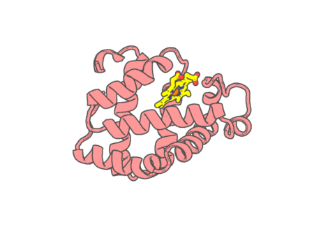 |
Experimental method | X-RAY DIFFRACTION |
|---|---|---|
| Structural data | available | |
| Non-standard amino acids | None | |
| Missing residues/ Structural gap | None / None | |
| Resolution | 1.81 Å | |
| Deposition date | 2010-12-02 |
| pdb_00003ark | Cl- binding hemoglobin component V form Propsilocerus akamusi under 1 M NaCl at pH 4.6 | ||
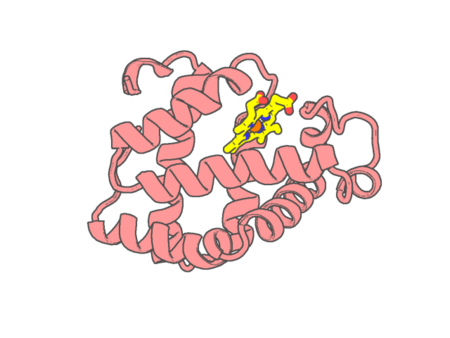 |
Experimental method | X-RAY DIFFRACTION |
|---|---|---|
| Structural data | available | |
| Non-standard amino acids | None | |
| Missing residues/ Structural gap | None / None | |
| Resolution | 1.81 Å | |
| Deposition date | 2010-12-02 |
| pdb_00003arl | Cl- binding hemoglobin component V form Propsilocerus akamusi under 500 mM NaCl at pH 5.5 | ||
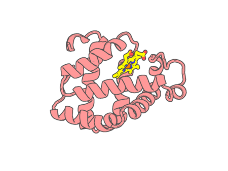 |
Experimental method | X-RAY DIFFRACTION |
|---|---|---|
| Structural data | available | |
| Non-standard amino acids | None | |
| Missing residues/ Structural gap | None / None | |
| Resolution | 1.81 Å | |
| Deposition date | 2010-12-02 |