
Number of data: 3440
Dataset: (without mutations)
| pdb_00006fwf | Low resolution structure of Neisseria meningitidis qNOR | ||
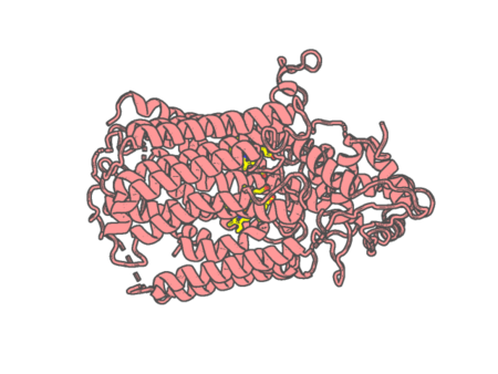 |
Experimental method | X-RAY DIFFRACTION |
|---|---|---|
| Structural data | available | |
| Non-standard amino acids | None | |
| Missing residues/ Structural gap | Exists / Exists | |
| Resolution | 4.2 Å | |
| Deposition date | 2018-03-06 |
| pdb_00006fyj | Cytochrome P450 peroxygenase CYP152K6 in complex with Myristic Acid | ||
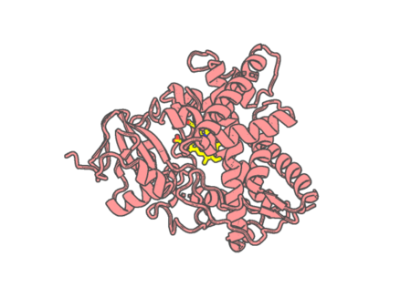 |
Experimental method | X-RAY DIFFRACTION |
|---|---|---|
| Structural data | available | |
| Non-standard amino acids | None | |
| Missing residues/ Structural gap | Exists / Exists | |
| Resolution | 1.3 Å | |
| Deposition date | 2018-03-12 |
| pdb_00006g5o | The structure of a carbohydrate active P450 | ||
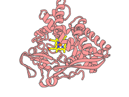 |
Experimental method | X-RAY DIFFRACTION |
|---|---|---|
| Structural data | available | |
| Non-standard amino acids | None | |
| Missing residues/ Structural gap | Exists / Exists | |
| Resolution | 2.25 Å | |
| Deposition date | 2018-03-29 |
| pdb_00006g5q | The structure of a carbohydrate active P450 | ||
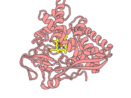 |
Experimental method | X-RAY DIFFRACTION |
|---|---|---|
| Structural data | available | |
| Non-standard amino acids | None | |
| Missing residues/ Structural gap | Exists / Exists | |
| Resolution | 2.4 Å | |
| Deposition date | 2018-03-29 |
| pdb_00006geo | Crystal structure of Mycobacterium tuberculosis cytochrome P450 CYP121A1 in complex with Triazole Pyrazole inhibitor 10j | ||
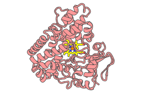 |
Experimental method | X-RAY DIFFRACTION |
|---|---|---|
| Structural data | available | |
| Non-standard amino acids | None | |
| Missing residues/ Structural gap | None / None | |
| Resolution | 1.5 Å | |
| Deposition date | 2018-04-27 |
| pdb_00006geq | Crystal structure of Mycobacterium tuberculosis cytochrome P450 CYP121A1 in complex with Triazole Pyrazole inhibitor 14a | ||
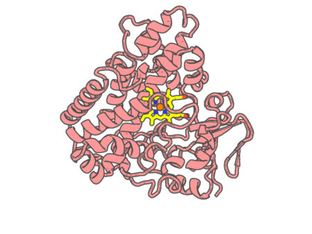 |
Experimental method | X-RAY DIFFRACTION |
|---|---|---|
| Structural data | available | |
| Non-standard amino acids | None | |
| Missing residues/ Structural gap | None / None | |
| Resolution | 1.6 Å | |
| Deposition date | 2018-04-27 |
| pdb_00006gii | The crystal structure of Tepidiphilus thermophilus P450 heme domain | ||
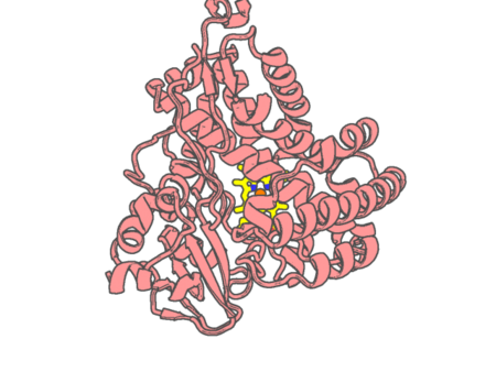 |
Experimental method | X-RAY DIFFRACTION |
|---|---|---|
| Structural data | available | |
| Non-standard amino acids | None | |
| Missing residues/ Structural gap | None / None | |
| Resolution | 1.9 Å | |
| Deposition date | 2018-05-11 |
| pdb_00006gk5 | Crystal structure of cytochrome P450 CYP267B1 from Sorangium cellulosum So ce56 | ||
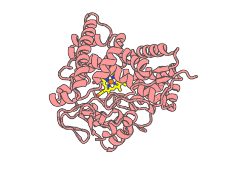 |
Experimental method | X-RAY DIFFRACTION |
|---|---|---|
| Structural data | available | |
| Non-standard amino acids | None | |
| Missing residues/ Structural gap | None / None | |
| Resolution | 1.6 Å | |
| Deposition date | 2018-05-18 |
| pdb_00006gk6 | Crystal structure of myxobacterial cytochrome P450 CYP267B1 in complex with myristic acid | ||
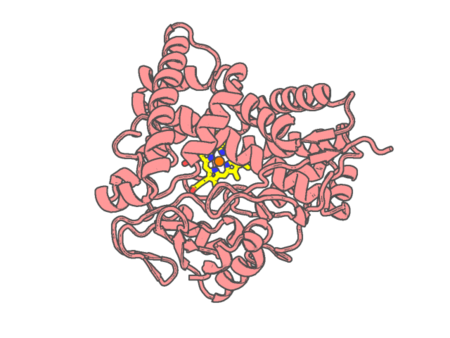 |
Experimental method | X-RAY DIFFRACTION |
|---|---|---|
| Structural data | available | |
| Non-standard amino acids | None | |
| Missing residues/ Structural gap | None / None | |
| Resolution | 1.6 Å | |
| Deposition date | 2018-05-18 |
| pdb_00006gmf | Structure of Cytochrome P450 CYP109Q5 from Chondromyces apiculatus | ||
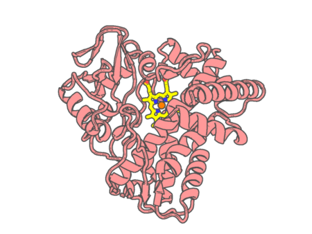 |
Experimental method | X-RAY DIFFRACTION |
|---|---|---|
| Structural data | available | |
| Non-standard amino acids | None | |
| Missing residues/ Structural gap | Exists / Exists | |
| Resolution | 1.55 Å | |
| Deposition date | 2018-05-25 |
| pdb_00006h5l | Kuenenia stuttgartiensis reducing HAO-like protein complex Kustc0457/Kustc0458 | ||
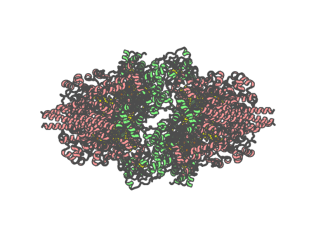 |
Experimental method | X-RAY DIFFRACTION |
|---|---|---|
| Structural data | available | |
| Non-standard amino acids | None | |
| Missing residues/ Structural gap | None / None | |
| Resolution | 2.6 Å | |
| Deposition date | 2018-07-25 |
| pdb_00006hal | Human carbonmonoxy hemoglobin SFX dataset | ||
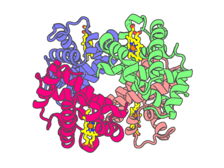 |
Experimental method | X-RAY DIFFRACTION |
|---|---|---|
| Structural data | available | |
| Non-standard amino acids | None | |
| Missing residues/ Structural gap | None / None | |
| Resolution | 2.2 Å | |
| Deposition date | 2018-08-07 |
| pdb_00006hcy | human STEAP4 bound to NADP, FAD, heme and Fe(III)-NTA. | ||
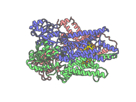 |
Experimental method | ELECTRON MICROSCOPY |
|---|---|---|
| Structural data | available | |
| Non-standard amino acids | None | |
| Missing residues/ Structural gap | None / None | |
| Resolution | None | |
| Deposition date | 2018-08-17 |
| pdb_00006hd1 | human STEAP4 bound to NADPH, FAD and heme. | ||
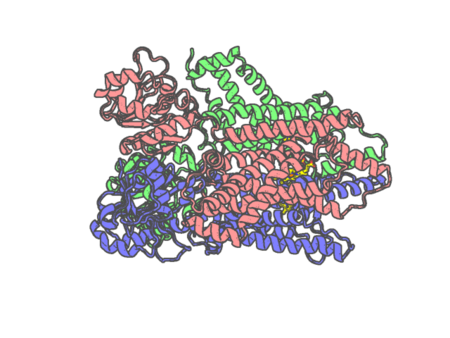 |
Experimental method | ELECTRON MICROSCOPY |
|---|---|---|
| Structural data | available | |
| Non-standard amino acids | None | |
| Missing residues/ Structural gap | None / None | |
| Resolution | None | |
| Deposition date | 2018-08-17 |
| pdb_00006hif | Kuenenia stuttgartiensis hydrazine dehydrogenase complex | ||
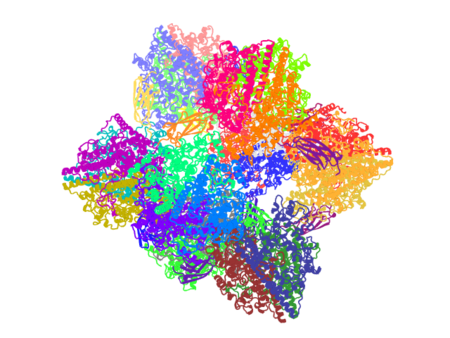 |
Experimental method | X-RAY DIFFRACTION |
|---|---|---|
| Structural data | available | |
| Non-standard amino acids | None | |
| Missing residues/ Structural gap | None / None | |
| Resolution | 2.8 Å | |
| Deposition date | 2018-08-29 |
| pdb_00006hih | Cytochrome c prime beta from Methylococcus capsulatus (Bath) | ||
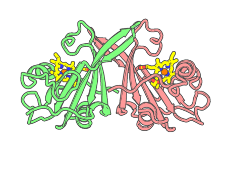 |
Experimental method | X-RAY DIFFRACTION |
|---|---|---|
| Structural data | available | |
| Non-standard amino acids | None | |
| Missing residues/ Structural gap | None / None | |
| Resolution | 1.6 Å | |
| Deposition date | 2018-08-29 |
| pdb_00006hiu | Cytochrome P460 from Methylococcus capsulatus (Bath) | ||
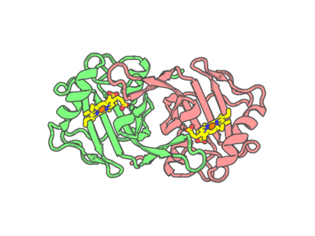 |
Experimental method | X-RAY DIFFRACTION |
|---|---|---|
| Structural data | available | |
| Non-standard amino acids | None | |
| Missing residues/ Structural gap | None / None | |
| Resolution | 1.36 Å | |
| Deposition date | 2018-08-31 |
| pdb_00006hk2 | Crystal structure of ferric R-state human methemoglobin bound to maleimide-deferoxamine bifunctional chelator (DFO) | ||
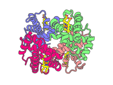 |
Experimental method | X-RAY DIFFRACTION |
|---|---|---|
| Structural data | available | |
| Non-standard amino acids | None | |
| Missing residues/ Structural gap | None / None | |
| Resolution | 1.55 Å | |
| Deposition date | 2018-09-05 |
| pdb_00006hqd | Cytochrome P450-153 from Pseudomonas sp. 19-rlim | ||
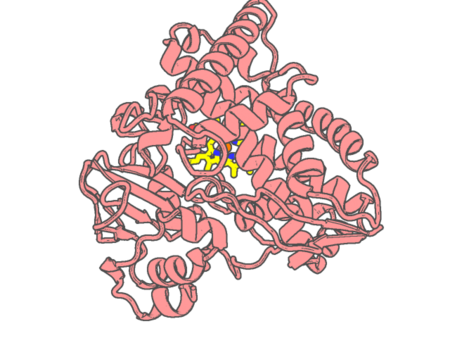 |
Experimental method | X-RAY DIFFRACTION |
|---|---|---|
| Structural data | available | |
| Non-standard amino acids | None | |
| Missing residues/ Structural gap | None / None | |
| Resolution | 1.8 Å | |
| Deposition date | 2018-09-24 |
| pdb_00006hqg | Cytochrome P450-153 from Phenylobacterium zucineum | ||
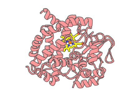 |
Experimental method | X-RAY DIFFRACTION |
|---|---|---|
| Structural data | unavailable | |
| Non-standard amino acids | None | |
| Missing residues/ Structural gap | Exists / Exists | |
| Resolution | 2.9 Å | |
| Deposition date | 2018-09-25 |