
Number of data: 3898
Dataset: (exptl_method: X-ray) and (resolution_limit=2.0)
| pdb_00005g6j | Structure of Bacillus subtilis Nitric Oxide Synthase in complex with 7-((3-(2-(Methylamino)ethyl)phenoxy)methyl)quinolin-2- amine | ||
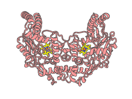 |
Experimental method | X-RAY DIFFRACTION |
|---|---|---|
| Structural data | available | |
| Non-standard amino acids | None | |
| Missing residues/ Structural gap | None / None | |
| Resolution | 1.88 Å | |
| Deposition date | 2016-06-18 |
| pdb_00005g6k | Structure of Bacillus subtilis Nitric Oxide Synthase I218V in complex with 7-((3-(2-(Methylamino)ethyl)phenoxy)methyl)quinolin-2- amine | ||
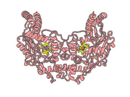 |
Experimental method | X-RAY DIFFRACTION |
|---|---|---|
| Structural data | available | |
| Non-standard amino acids | None | |
| Missing residues/ Structural gap | None / None | |
| Resolution | 1.951 Å | |
| Deposition date | 2016-06-18 |
| pdb_00005g6m | Structure of Bacillus subtilis Nitric Oxide Synthase in complex with 7-((3-Aminomethyl)phenoxy)methyl)quinolin-2-amine | ||
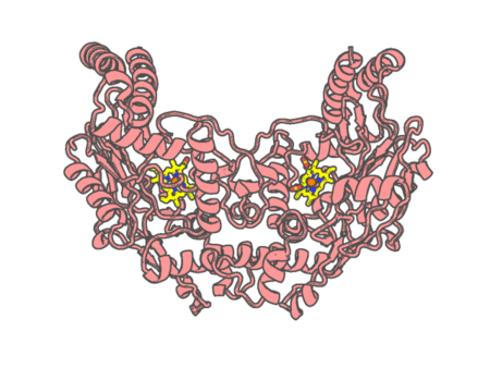 |
Experimental method | X-RAY DIFFRACTION |
|---|---|---|
| Structural data | available | |
| Non-standard amino acids | None | |
| Missing residues/ Structural gap | None / None | |
| Resolution | 1.77 Å | |
| Deposition date | 2016-06-18 |
| pdb_00005g6n | Structure of Bacillus subtilis Nitric Oxide Synthase in complex with 7-((4-(Dimethylamino)methyl)phenoxy)methyl)quinolin-2- amine | ||
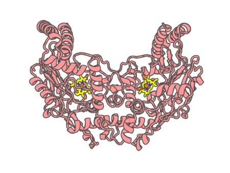 |
Experimental method | X-RAY DIFFRACTION |
|---|---|---|
| Structural data | available | |
| Non-standard amino acids | None | |
| Missing residues/ Structural gap | None / None | |
| Resolution | 1.91 Å | |
| Deposition date | 2016-06-18 |
| pdb_00005g6o | Structure of Bacillus subtilis Nitric Oxide Synthase in complex with two molecules of 7-((4-(Dimethylamino)methyl)phenoxy)methyl)quinolin- 2- amine | ||
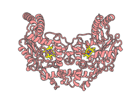 |
Experimental method | X-RAY DIFFRACTION |
|---|---|---|
| Structural data | available | |
| Non-standard amino acids | None | |
| Missing residues/ Structural gap | None / None | |
| Resolution | 1.719 Å | |
| Deposition date | 2016-06-18 |
| pdb_00005gj3 | Periplasmic heme-binding protein RhuT from Roseiflexus sp. RS-1 in two-heme bound form (holo-2) | ||
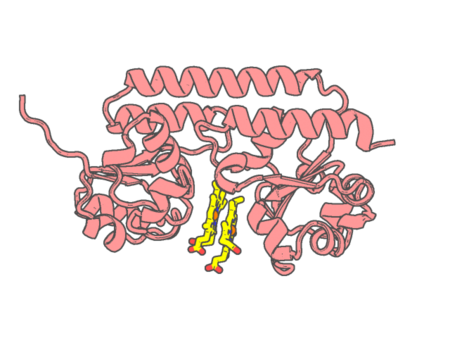 |
Experimental method | X-RAY DIFFRACTION |
|---|---|---|
| Structural data | available | |
| Non-standard amino acids | None | |
| Missing residues/ Structural gap | None / None | |
| Resolution | 2 Å | |
| Deposition date | 2016-06-26 |
| pdb_00005gls | Structure of bovine Lactoperoxidase with a partially modified covalent bond with heme moiety | ||
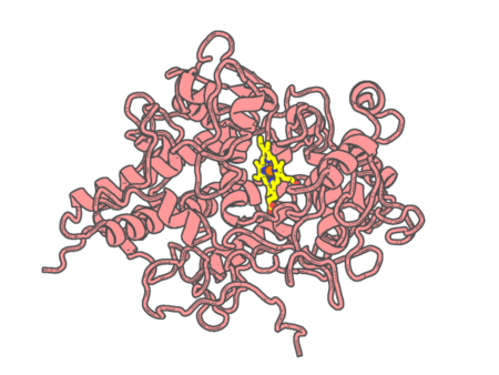 |
Experimental method | X-RAY DIFFRACTION |
|---|---|---|
| Structural data | available | |
| Non-standard amino acids | Included | |
| Missing residues/ Structural gap | None / None | |
| Resolution | 1.93 Å | |
| Deposition date | 2016-07-12 |
| pdb_00005gnl | Cytochrome P450 Vdh (CYP107BR1) F106V mutant | ||
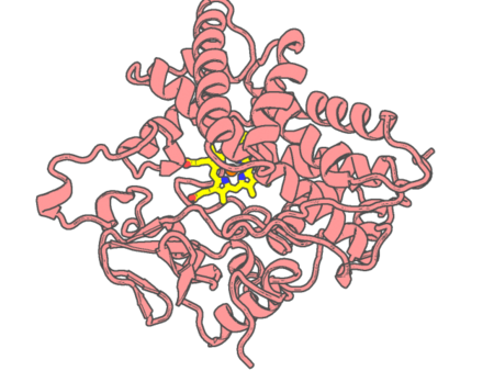 |
Experimental method | X-RAY DIFFRACTION |
|---|---|---|
| Structural data | available | |
| Non-standard amino acids | None | |
| Missing residues/ Structural gap | None / None | |
| Resolution | 1.95 Å | |
| Deposition date | 2016-07-22 |
| pdb_00005gwe | cytochrome P450 CREJ | ||
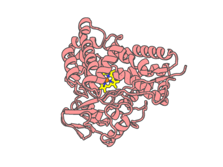 |
Experimental method | X-RAY DIFFRACTION |
|---|---|---|
| Structural data | available | |
| Non-standard amino acids | None | |
| Missing residues/ Structural gap | None / None | |
| Resolution | 2 Å | |
| Deposition date | 2016-09-11 |
| pdb_00005gxg | High-resolution crystal structure of the electron transfer complex of cytochrome p450cam with putidaredoxin | ||
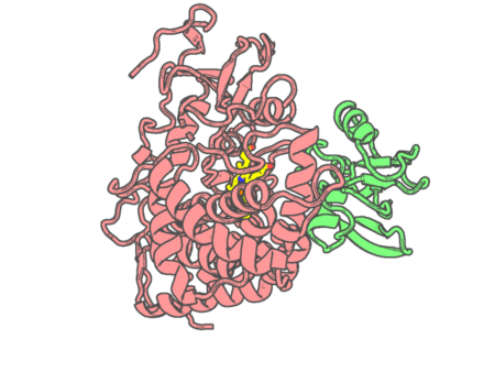 |
Experimental method | X-RAY DIFFRACTION |
|---|---|---|
| Structural data | available | |
| Non-standard amino acids | None | |
| Missing residues/ Structural gap | Exists / Exists | |
| Resolution | 1.7 Å | |
| Deposition date | 2016-09-17 |
| pdb_00005gyr | Tetrameric Allochromatium vinosum cytochrome c' | ||
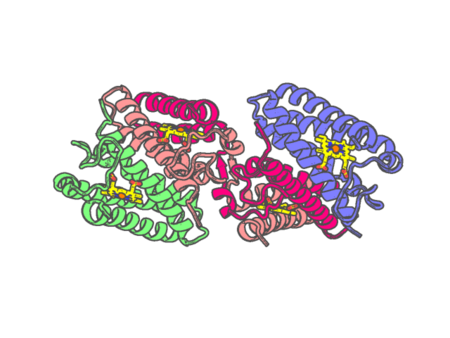 |
Experimental method | X-RAY DIFFRACTION |
|---|---|---|
| Structural data | available | |
| Non-standard amino acids | None | |
| Missing residues/ Structural gap | None / None | |
| Resolution | 1.6 Å | |
| Deposition date | 2016-09-23 |
| pdb_00005hav | Sperm whale myoglobin mutant L29H F33Y F43H (F33Y CuBMb) with oxygen bound | ||
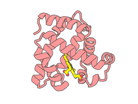 |
Experimental method | X-RAY DIFFRACTION |
|---|---|---|
| Structural data | available | |
| Non-standard amino acids | None | |
| Missing residues/ Structural gap | None / None | |
| Resolution | 1.268 Å | |
| Deposition date | 2015-12-31 |
| pdb_00005hbi | SCAPHARCA DIMERIC HEMOGLOBIN, MUTANT T72I, CO-LIGANDED FORM | ||
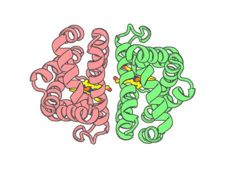 |
Experimental method | X-RAY DIFFRACTION |
|---|---|---|
| Structural data | available | |
| Non-standard amino acids | None | |
| Missing residues/ Structural gap | None / None | |
| Resolution | 1.6 Å | |
| Deposition date | 1998-06-24 |
| pdb_00005hdi | Structural characterization of CYP144A1, a Mycobacterium tuberculosis cytochrome P450 | ||
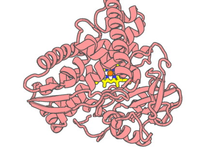 |
Experimental method | X-RAY DIFFRACTION |
|---|---|---|
| Structural data | available | |
| Non-standard amino acids | None | |
| Missing residues/ Structural gap | Exists / Exists | |
| Resolution | 1.54 Å | |
| Deposition date | 2016-01-05 |
| pdb_00005hiw | Sorangium cellulosum So Ce56 cytochrome P450 260B1 | ||
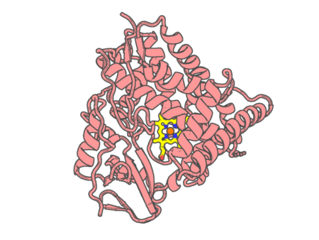 |
Experimental method | X-RAY DIFFRACTION |
|---|---|---|
| Structural data | available | |
| Non-standard amino acids | None | |
| Missing residues/ Structural gap | Exists / Exists | |
| Resolution | 1.85 Å | |
| Deposition date | 2016-01-12 |
| pdb_00005hlq | X-ray crystal structure of met F43H/H64A sperm whale myoglobin | ||
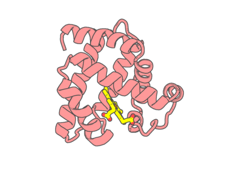 |
Experimental method | X-RAY DIFFRACTION |
|---|---|---|
| Structural data | available | |
| Non-standard amino acids | None | |
| Missing residues/ Structural gap | None / None | |
| Resolution | 1.5 Å | |
| Deposition date | 2016-01-15 |
| pdb_00005hlu | X-ray crystal structure of met F43H/H64A sperm whale myoglobin in complex with nitric oxide | ||
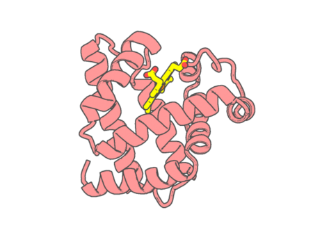 |
Experimental method | X-RAY DIFFRACTION |
|---|---|---|
| Structural data | available | |
| Non-standard amino acids | None | |
| Missing residues/ Structural gap | None / None | |
| Resolution | 1.5 Å | |
| Deposition date | 2016-01-15 |
| pdb_00005hlx | X-ray crystal structure of met F43H/H64A sperm whale myoglobin in complex with nitrite | ||
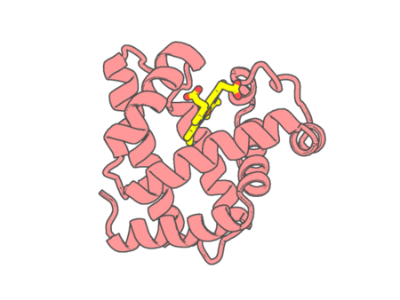 |
Experimental method | X-RAY DIFFRACTION |
|---|---|---|
| Structural data | available | |
| Non-standard amino acids | None | |
| Missing residues/ Structural gap | None / None | |
| Resolution | 1.5 Å | |
| Deposition date | 2016-01-15 |
| pdb_00005hwz | Crystal structure of nitrophorin 4 D30N mutant with nitrite | ||
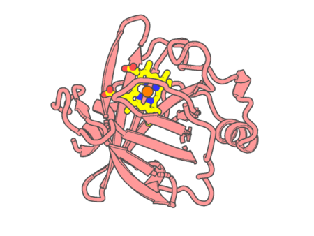 |
Experimental method | X-RAY DIFFRACTION |
|---|---|---|
| Structural data | available | |
| Non-standard amino acids | None | |
| Missing residues/ Structural gap | None / None | |
| Resolution | 1.45 Å | |
| Deposition date | 2016-01-29 |
| pdb_00005ibd | Crystal structure Mycobacterium tuberculosis CYP121 in complex with inhibitor fragment 24a | ||
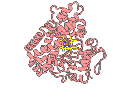 |
Experimental method | X-RAY DIFFRACTION |
|---|---|---|
| Structural data | available | |
| Non-standard amino acids | None | |
| Missing residues/ Structural gap | None / None | |
| Resolution | 1.77 Å | |
| Deposition date | 2016-02-22 |