
Number of data: 3898
Dataset: (exptl_method: X-ray) and (resolution_limit=2.0)
| pdb_00005lft | Crystal structure of cytochrome c - Bromo-trisulfonatocalix[4]arene complexes | ||
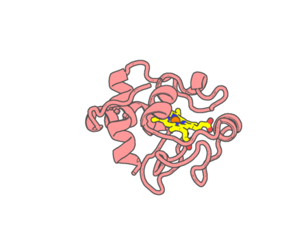 |
Experimental method | X-RAY DIFFRACTION |
|---|---|---|
| Structural data | available | |
| Non-standard amino acids | None | |
| Missing residues/ Structural gap | None / None | |
| Resolution | 1.249 Å | |
| Deposition date | 2016-07-04 |
| pdb_00005li6 | Crystal structure of Mycobacterium tuberculosis CYP126A1 in complex with N-isopropyl-N-((3-(4-methoxyphenyl)-1,2,4-oxadiazol-5-yl)methyl)-2-(4-nitrophenyl)acetamide | ||
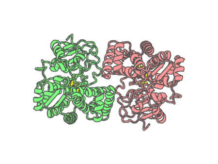 |
Experimental method | X-RAY DIFFRACTION |
|---|---|---|
| Structural data | unavailable | |
| Non-standard amino acids | None | |
| Missing residues/ Structural gap | Exists / Exists | |
| Resolution | 1.95 Å | |
| Deposition date | 2016-07-14 |
| pdb_00005li7 | Crystal structure of Mycobacterium tuberculosis CYP126A1 in complex with 1-(3-(1H-imidazol-1-yl)propyl)-3-((3s,5s,7s)-adamantan-1-yl)urea | ||
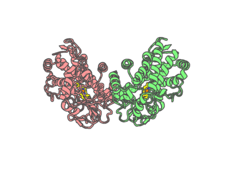 |
Experimental method | X-RAY DIFFRACTION |
|---|---|---|
| Structural data | unavailable | |
| Non-standard amino acids | None | |
| Missing residues/ Structural gap | Exists / Exists | |
| Resolution | 1.58 Å | |
| Deposition date | 2016-07-14 |
| pdb_00005li8 | Crystal structure of Mycobacterium tuberculosis CYP126A1 in complex with ketoconazole | ||
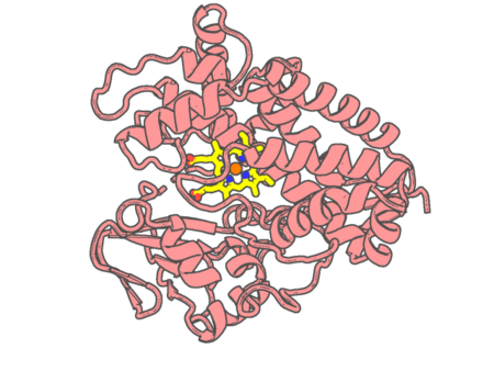 |
Experimental method | X-RAY DIFFRACTION |
|---|---|---|
| Structural data | available | |
| Non-standard amino acids | None | |
| Missing residues/ Structural gap | Exists / Exists | |
| Resolution | 1.83 Å | |
| Deposition date | 2016-07-14 |
| pdb_00005lie | Crystal structure of Mycobacterium tuberculosis CYP126A1 in complex with imidazole | ||
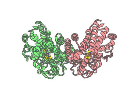 |
Experimental method | X-RAY DIFFRACTION |
|---|---|---|
| Structural data | available | |
| Non-standard amino acids | None | |
| Missing residues/ Structural gap | Exists / Exists | |
| Resolution | 1.799 Å | |
| Deposition date | 2016-07-14 |
| pdb_00005lk9 | Dehaloperoxidase B from Amphitrite ornata: indazole complex | ||
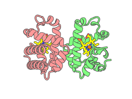 |
Experimental method | X-RAY DIFFRACTION |
|---|---|---|
| Structural data | available | |
| Non-standard amino acids | None | |
| Missing residues/ Structural gap | None / None | |
| Resolution | 1.12 Å | |
| Deposition date | 2016-07-21 |
| pdb_00005lkv | Dehaloperoxidase B from Amphitrite ornata: imidazole complex | ||
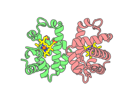 |
Experimental method | X-RAY DIFFRACTION |
|---|---|---|
| Structural data | available | |
| Non-standard amino acids | None | |
| Missing residues/ Structural gap | None / None | |
| Resolution | 1.08 Å | |
| Deposition date | 2016-07-25 |
| pdb_00005llz | Dehaloperoxidase B from Amphitrite ornata: benzotriazole complex | ||
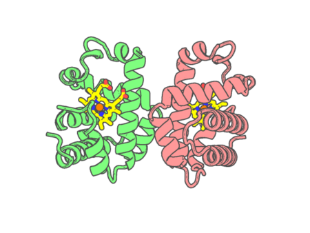 |
Experimental method | X-RAY DIFFRACTION |
|---|---|---|
| Structural data | available | |
| Non-standard amino acids | None | |
| Missing residues/ Structural gap | None / None | |
| Resolution | 1.14 Å | |
| Deposition date | 2016-07-28 |
| pdb_00005lte | Crystal structure of the alpha subunit of heme dependent oxidative N-demethylase (HODM) | ||
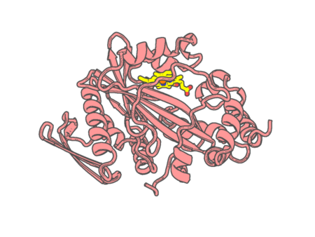 |
Experimental method | X-RAY DIFFRACTION |
|---|---|---|
| Structural data | available | |
| Non-standard amino acids | None | |
| Missing residues/ Structural gap | None / None | |
| Resolution | 1.65 Å | |
| Deposition date | 2016-09-06 |
| pdb_00005lth | Crystal structure of the alpha subunit of heme dependent oxidative N-demethylase (HODM) in complex with the dimethylamine substrate | ||
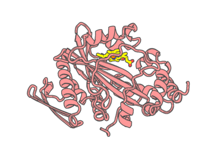 |
Experimental method | X-RAY DIFFRACTION |
|---|---|---|
| Structural data | available | |
| Non-standard amino acids | None | |
| Missing residues/ Structural gap | None / None | |
| Resolution | 1.76 Å | |
| Deposition date | 2016-09-06 |
| pdb_00005lti | Crystal structure of the alpha subunit of heme dependent oxidative N-demethylase (HODM) in complex with the dimethylamine substrate | ||
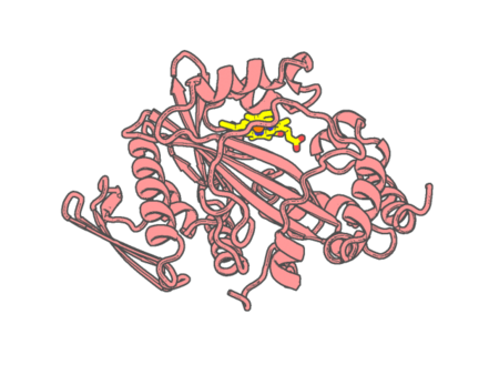 |
Experimental method | X-RAY DIFFRACTION |
|---|---|---|
| Structural data | available | |
| Non-standard amino acids | None | |
| Missing residues/ Structural gap | None / None | |
| Resolution | 1.9 Å | |
| Deposition date | 2016-09-06 |
| pdb_00005lyc | Cytochrome c in complex with phosphonato-calix[6]arene | ||
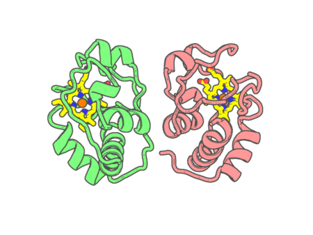 |
Experimental method | X-RAY DIFFRACTION |
|---|---|---|
| Structural data | available | |
| Non-standard amino acids | None | |
| Missing residues/ Structural gap | None / None | |
| Resolution | 1.8 Å | |
| Deposition date | 2016-09-27 |
| pdb_00005m0n | Crystal structure of cytochrome P450 OleT in complex with formate | ||
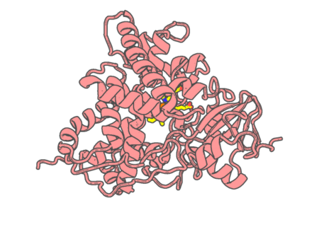 |
Experimental method | X-RAY DIFFRACTION |
|---|---|---|
| Structural data | available | |
| Non-standard amino acids | None | |
| Missing residues/ Structural gap | None / None | |
| Resolution | 1.44 Å | |
| Deposition date | 2016-10-05 |
| pdb_00005m0o | Crystal structure of cytochrome P450 OleT H85Q in complex with arachidonic acid | ||
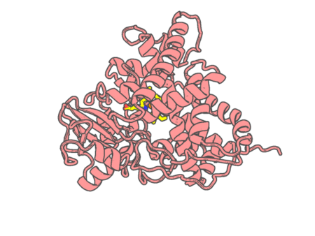 |
Experimental method | X-RAY DIFFRACTION |
|---|---|---|
| Structural data | available | |
| Non-standard amino acids | None | |
| Missing residues/ Structural gap | None / None | |
| Resolution | 1.8 Å | |
| Deposition date | 2016-10-05 |
| pdb_00005m0p | Crystal structure of cytochrome P450 OleT F79A in complex with arachidonic acid | ||
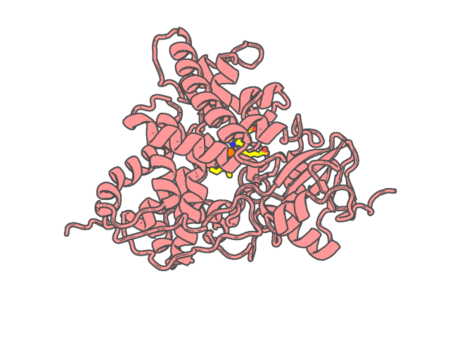 |
Experimental method | X-RAY DIFFRACTION |
|---|---|---|
| Structural data | available | |
| Non-standard amino acids | None | |
| Missing residues/ Structural gap | None / None | |
| Resolution | 1.95 Å | |
| Deposition date | 2016-10-05 |
| pdb_00005m3s | Low-dose fixed target serial synchrotron crystallography structure of Metmyoglobin | ||
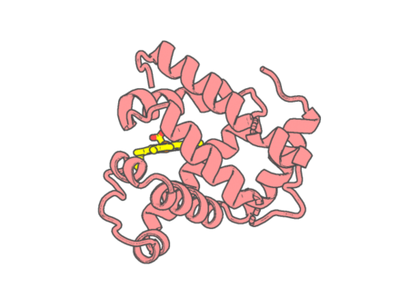 |
Experimental method | X-RAY DIFFRACTION |
|---|---|---|
| Structural data | available | |
| Non-standard amino acids | None | |
| Missing residues/ Structural gap | None / None | |
| Resolution | 1.8 Å | |
| Deposition date | 2016-10-17 |
| pdb_00005m6j | Crystal structure of nitrophorin 7 E27V mutant from Rhodnius prolixus | ||
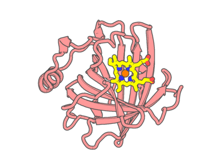 |
Experimental method | X-RAY DIFFRACTION |
|---|---|---|
| Structural data | available | |
| Non-standard amino acids | None | |
| Missing residues/ Structural gap | None / None | |
| Resolution | 1.7 Å | |
| Deposition date | 2016-10-25 |
| pdb_00005m6k | Crystal structure of nitrophorin 7 E27V mutant from Rhodnius prolixus with imidazole | ||
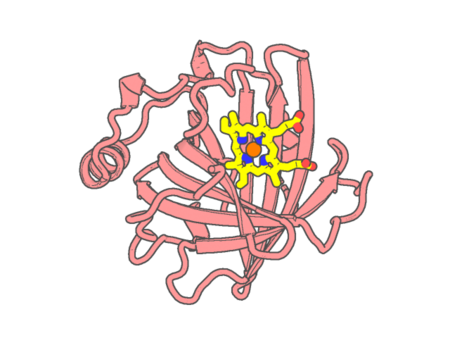 |
Experimental method | X-RAY DIFFRACTION |
|---|---|---|
| Structural data | available | |
| Non-standard amino acids | None | |
| Missing residues/ Structural gap | None / None | |
| Resolution | 1.6 Å | |
| Deposition date | 2016-10-25 |
| pdb_00005map | X-ray generated oxyferrous complex of DtpA from Streptomyces lividans | ||
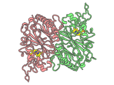 |
Experimental method | X-RAY DIFFRACTION |
|---|---|---|
| Structural data | available | |
| Non-standard amino acids | None | |
| Missing residues/ Structural gap | Exists / Exists | |
| Resolution | 1.49 Å | |
| Deposition date | 2016-11-04 |
| pdb_00005mau | Crystal structure of dimeric chlorite dismutase from Cyanothece sp. PCC7425 (pH 6.5) | ||
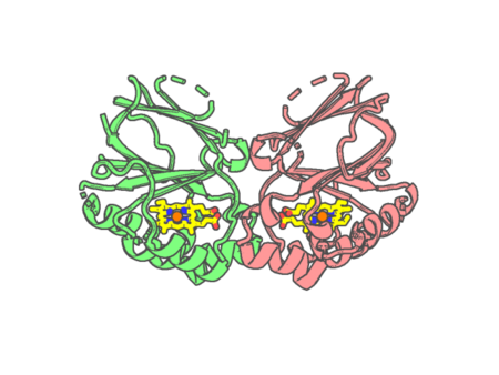 |
Experimental method | X-RAY DIFFRACTION |
|---|---|---|
| Structural data | available | |
| Non-standard amino acids | None | |
| Missing residues/ Structural gap | Exists / Exists | |
| Resolution | 1.3 Å | |
| Deposition date | 2016-11-04 |