
Number of data: 3898
Dataset: (exptl_method: X-ray) and (resolution_limit=2.0)
| pdb_00005o1l | Structure of Latex Clearing Protein LCP in the open state with bound imidazole | ||
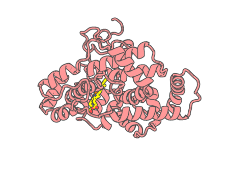 |
Experimental method | X-RAY DIFFRACTION |
|---|---|---|
| Structural data | available | |
| Non-standard amino acids | None | |
| Missing residues/ Structural gap | None / None | |
| Resolution | 1.48 Å | |
| Deposition date | 2017-05-18 |
| pdb_00005o41 | Low-dose fixed target serial synchrotron crystallography structure of sperm whale myoglobin | ||
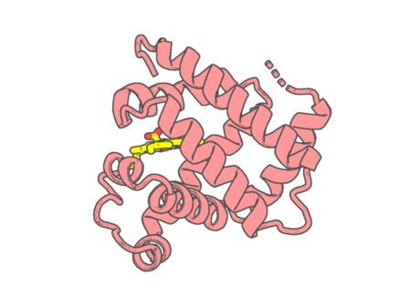 |
Experimental method | X-RAY DIFFRACTION |
|---|---|---|
| Structural data | available | |
| Non-standard amino acids | None | |
| Missing residues/ Structural gap | None / None | |
| Resolution | 1.8 Å | |
| Deposition date | 2017-05-25 |
| pdb_00005o4k | Crystal structure of P450 CYP121 in complex with compound 6b. | ||
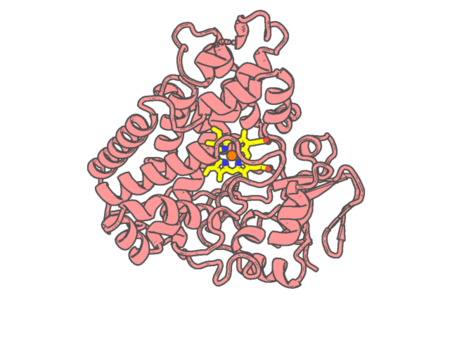 |
Experimental method | X-RAY DIFFRACTION |
|---|---|---|
| Structural data | available | |
| Non-standard amino acids | None | |
| Missing residues/ Structural gap | None / None | |
| Resolution | 1.5 Å | |
| Deposition date | 2017-05-29 |
| pdb_00005o4l | Crystal structure of P450 CYP121 in complex with compound 6a. | ||
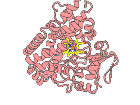 |
Experimental method | X-RAY DIFFRACTION |
|---|---|---|
| Structural data | available | |
| Non-standard amino acids | None | |
| Missing residues/ Structural gap | None / None | |
| Resolution | 1.64 Å | |
| Deposition date | 2017-05-29 |
| pdb_00005obo | Crystal structure of nitrite bound D97N mutant of three-domain heme-Cu nitrite reductase from Ralstonia pickettii | ||
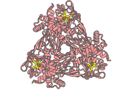 |
Experimental method | X-RAY DIFFRACTION |
|---|---|---|
| Structural data | available | |
| Non-standard amino acids | None | |
| Missing residues/ Structural gap | None / None | |
| Resolution | 1.89 Å | |
| Deposition date | 2017-06-28 |
| pdb_00005oc0 | Structure of E. coli superoxide oxidase | ||
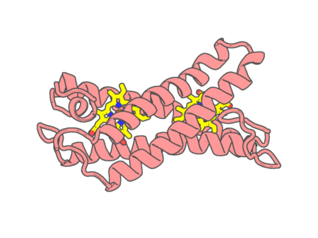 |
Experimental method | X-RAY DIFFRACTION |
|---|---|---|
| Structural data | available | |
| Non-standard amino acids | Included | |
| Missing residues/ Structural gap | None / None | |
| Resolution | 1.97 Å | |
| Deposition date | 2017-06-29 |
| pdb_00005ocb | Crystal structure of nitric oxide bound D97N mutant of three-domain heme-Cu nitrite reductase from Ralstonia pickettii | ||
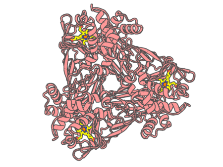 |
Experimental method | X-RAY DIFFRACTION |
|---|---|---|
| Structural data | available | |
| Non-standard amino acids | None | |
| Missing residues/ Structural gap | None / None | |
| Resolution | 1.78 Å | |
| Deposition date | 2017-06-30 |
| pdb_00005ocf | Crystal structure of nitric oxide bound to three-domain heme-Cu nitrite reductase from Ralstonia pickettii | ||
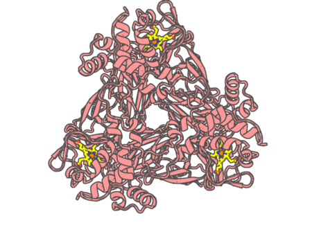 |
Experimental method | X-RAY DIFFRACTION |
|---|---|---|
| Structural data | available | |
| Non-standard amino acids | None | |
| Missing residues/ Structural gap | None / None | |
| Resolution | 1.8 Å | |
| Deposition date | 2017-06-30 |
| pdb_00005ohe | Globin sensor domain of AfGcHK (FeIII form) in complex with cyanide | ||
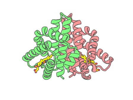 |
Experimental method | X-RAY DIFFRACTION |
|---|---|---|
| Structural data | available | |
| Non-standard amino acids | None | |
| Missing residues/ Structural gap | None / None | |
| Resolution | 1.85 Å | |
| Deposition date | 2017-07-16 |
| pdb_00005ohf | Globin sensor domain of AfGcHK (FeIII form) in complex with cyanide, partially reduced | ||
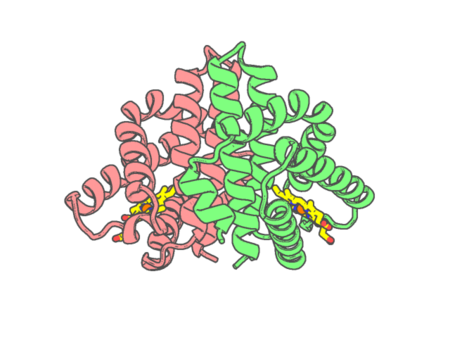 |
Experimental method | X-RAY DIFFRACTION |
|---|---|---|
| Structural data | available | |
| Non-standard amino acids | None | |
| Missing residues/ Structural gap | None / None | |
| Resolution | 1.8 Å | |
| Deposition date | 2017-07-16 |
| pdb_00005oj9 | Structure of Mb NMH | ||
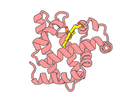 |
Experimental method | X-RAY DIFFRACTION |
|---|---|---|
| Structural data | available | |
| Non-standard amino acids | Included | |
| Missing residues/ Structural gap | None / None | |
| Resolution | 1.483 Å | |
| Deposition date | 2017-07-21 |
| pdb_00005oja | Structure of MbQ | ||
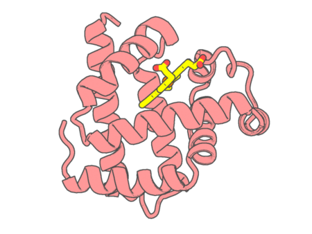 |
Experimental method | X-RAY DIFFRACTION |
|---|---|---|
| Structural data | available | |
| Non-standard amino acids | None | |
| Missing residues/ Structural gap | None / None | |
| Resolution | 1.347 Å | |
| Deposition date | 2017-07-21 |
| pdb_00005ojb | Structure of MbQ NMH | ||
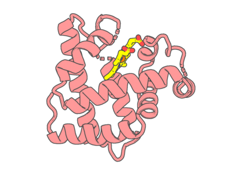 |
Experimental method | X-RAY DIFFRACTION |
|---|---|---|
| Structural data | available | |
| Non-standard amino acids | Included | |
| Missing residues/ Structural gap | None / None | |
| Resolution | 1.543 Å | |
| Deposition date | 2017-07-21 |
| pdb_00005ojc | Structure of MbQ2.1 NMH | ||
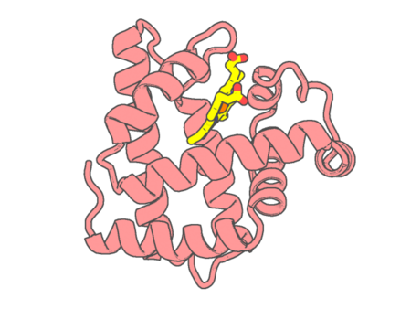 |
Experimental method | X-RAY DIFFRACTION |
|---|---|---|
| Structural data | available | |
| Non-standard amino acids | Included | |
| Missing residues/ Structural gap | None / None | |
| Resolution | 1.25 Å | |
| Deposition date | 2017-07-21 |
| pdb_00005omr | Crystal structure of Amycolatopsis cytochrome P450 GcoA in complex with vanillin. | ||
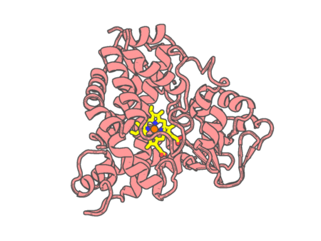 |
Experimental method | X-RAY DIFFRACTION |
|---|---|---|
| Structural data | available | |
| Non-standard amino acids | None | |
| Missing residues/ Structural gap | None / None | |
| Resolution | 1.68 Å | |
| Deposition date | 2017-08-01 |
| pdb_00005oms | Crystal structure of Amycolatopsis cytochrome P450 GcoA in complex with guaethol. | ||
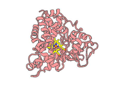 |
Experimental method | X-RAY DIFFRACTION |
|---|---|---|
| Structural data | available | |
| Non-standard amino acids | None | |
| Missing residues/ Structural gap | None / None | |
| Resolution | 1.95 Å | |
| Deposition date | 2017-08-01 |
| pdb_00005omu | Crystal structure of Amycolatopsis cytochrome P450 GcoA in complex with syringol | ||
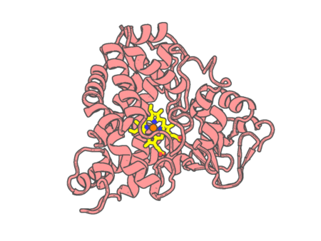 |
Experimental method | X-RAY DIFFRACTION |
|---|---|---|
| Structural data | available | |
| Non-standard amino acids | None | |
| Missing residues/ Structural gap | None / None | |
| Resolution | 1.95 Å | |
| Deposition date | 2017-08-01 |
| pdb_00005op9 | The crystal structure of P450 CYP121 in complex with lead compound 7e | ||
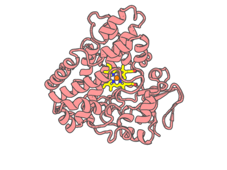 |
Experimental method | X-RAY DIFFRACTION |
|---|---|---|
| Structural data | available | |
| Non-standard amino acids | None | |
| Missing residues/ Structural gap | None / None | |
| Resolution | 1.455 Å | |
| Deposition date | 2017-08-09 |
| pdb_00005opa | The crystal structure of P450 CYP121 in complex with lead compound 7b | ||
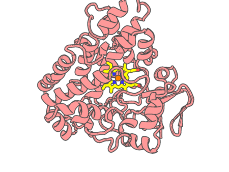 |
Experimental method | X-RAY DIFFRACTION |
|---|---|---|
| Structural data | available | |
| Non-standard amino acids | None | |
| Missing residues/ Structural gap | None / None | |
| Resolution | 1.345 Å | |
| Deposition date | 2017-08-09 |
| pdb_00005oxt | DIRECT-EVOLUTIONED UNSPECIFIC PEROXYGENASE FROM AGROCYBE AEGERITA, IN COMPLEX WITH ACETATE | ||
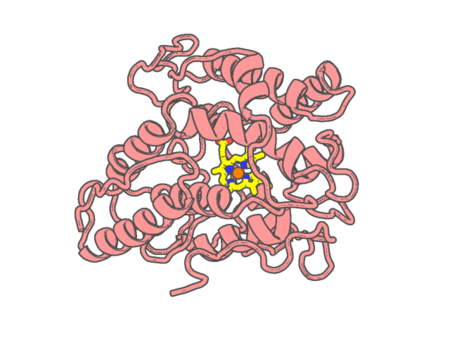 |
Experimental method | X-RAY DIFFRACTION |
|---|---|---|
| Structural data | available | |
| Non-standard amino acids | None | |
| Missing residues/ Structural gap | None / None | |
| Resolution | 1.42 Å | |
| Deposition date | 2017-09-07 |