
Number of data: 7660
Dataset: all data
| pdb_00003h34 | PpcE, A cytochrome c7 from Geobacter sulfurreducens | ||
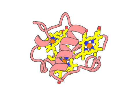 |
Experimental method | X-RAY DIFFRACTION |
|---|---|---|
| Structural data | available | |
| Non-standard amino acids | None | |
| Missing residues/ Structural gap | Exists / None | |
| Resolution | 1.6 Å | |
| Deposition date | 2009-04-15 |
| pdb_00003h4n | PpcD, A cytochrome c7 from Geobacter sulfurreducens | ||
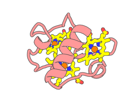 |
Experimental method | X-RAY DIFFRACTION |
|---|---|---|
| Structural data | available | |
| Non-standard amino acids | None | |
| Missing residues/ Structural gap | Exists / None | |
| Resolution | 1.35 Å | |
| Deposition date | 2009-04-20 |
| pdb_00003h57 | Myoglobin Cavity Mutant H64LV68N Deoxy form | ||
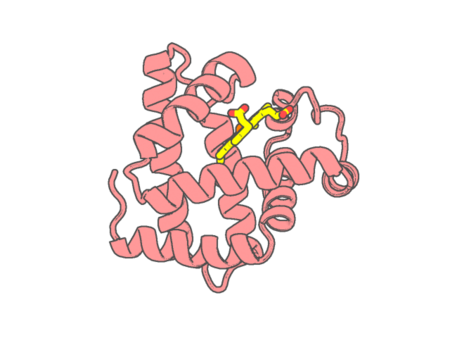 |
Experimental method | X-RAY DIFFRACTION |
|---|---|---|
| Structural data | available | |
| Non-standard amino acids | None | |
| Missing residues/ Structural gap | None / None | |
| Resolution | 1.7 Å | |
| Deposition date | 2009-04-21 |
| pdb_00003h58 | Myoglobin Cavity Mutant H64LV68N Met form | ||
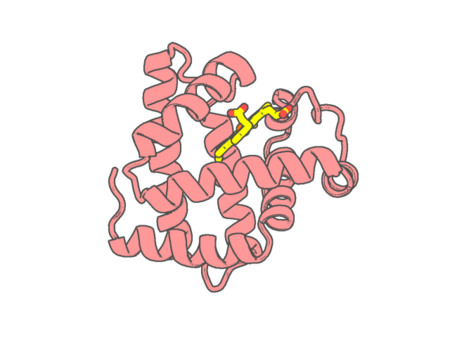 |
Experimental method | X-RAY DIFFRACTION |
|---|---|---|
| Structural data | available | |
| Non-standard amino acids | None | |
| Missing residues/ Structural gap | None / None | |
| Resolution | 1.8 Å | |
| Deposition date | 2009-04-21 |
| pdb_00003h8t | Structure of Porphyromonas gingivalis heme-binding protein HmuY in complex with Heme | ||
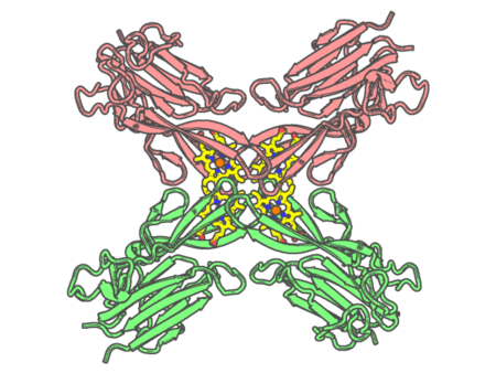 |
Experimental method | X-RAY DIFFRACTION |
|---|---|---|
| Structural data | available | |
| Non-standard amino acids | None | |
| Missing residues/ Structural gap | None / None | |
| Resolution | 1.8 Å | |
| Deposition date | 2009-04-29 |
| pdb_00003hb3 | High resolution crystal structure of Paracoccus denitrificans cytochrome c oxidase | ||
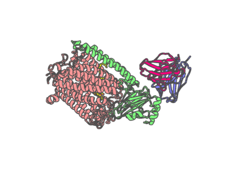 |
Experimental method | X-RAY DIFFRACTION |
|---|---|---|
| Structural data | available | |
| Non-standard amino acids | None | |
| Missing residues/ Structural gap | None / None | |
| Resolution | 2.25 Å | |
| Deposition date | 2009-05-04 |
| pdb_00003hb6 | Inactive mutant H54F of Proteus mirabilis catalase | ||
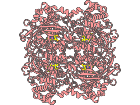 |
Experimental method | X-RAY DIFFRACTION |
|---|---|---|
| Structural data | available | |
| Non-standard amino acids | None | |
| Missing residues/ Structural gap | None / None | |
| Resolution | 2.3 Å | |
| Deposition date | 2009-05-04 |
| pdb_00003hc9 | Ferric Horse Heart Myoglobin; H64V mutant | ||
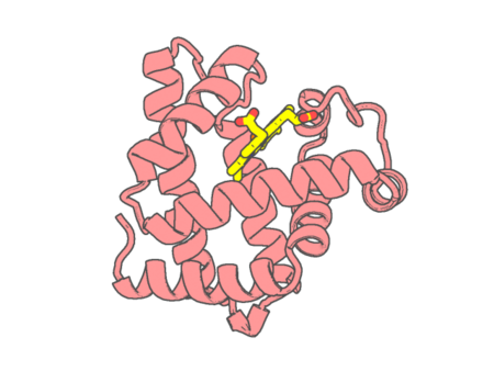 |
Experimental method | X-RAY DIFFRACTION |
|---|---|---|
| Structural data | available | |
| Non-standard amino acids | None | |
| Missing residues/ Structural gap | None / None | |
| Resolution | 2 Å | |
| Deposition date | 2009-05-05 |
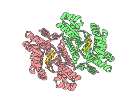
| pdb_00003hcn | Hg and protoporphyrin bound Human Ferrochelatase | ||
 |
Experimental method | X-RAY DIFFRACTION |
|---|---|---|
| Structural data | available | |
| Non-standard amino acids | None | |
| Missing residues/ Structural gap | None / None | |
| Resolution | 1.6 Å | |
| Deposition date | 2009-05-06 |
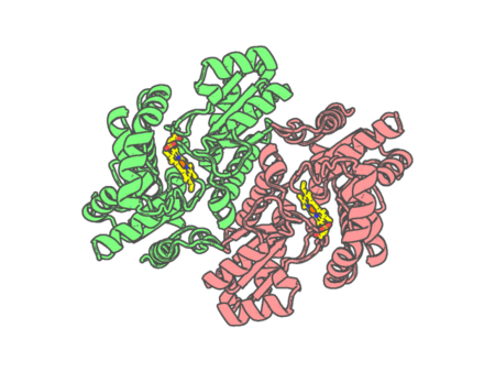
| pdb_00003hco | Human ferrochelatase with Cd and protoporphyrin IX bound | ||
 |
Experimental method | X-RAY DIFFRACTION |
|---|---|---|
| Structural data | available | |
| Non-standard amino acids | None | |
| Missing residues/ Structural gap | None / None | |
| Resolution | 1.8 Å | |
| Deposition date | 2009-05-06 |
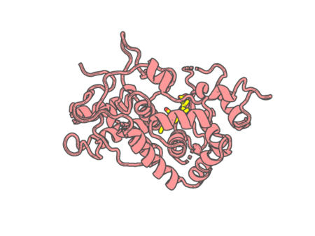
| pdb_00003hdl | Crystal Structure of Highly Glycosylated Peroxidase from Royal Palm Tree | ||
 |
Experimental method | X-RAY DIFFRACTION |
|---|---|---|
| Structural data | available | |
| Non-standard amino acids | None | |
| Missing residues/ Structural gap | None / None | |
| Resolution | 1.85 Å | |
| Deposition date | 2009-05-07 |
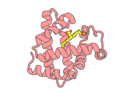
| pdb_00003hen | Ferric Horse Heart Myoglobin; H64V/V67R Mutant | ||
 |
Experimental method | X-RAY DIFFRACTION |
|---|---|---|
| Structural data | available | |
| Non-standard amino acids | None | |
| Missing residues/ Structural gap | None / None | |
| Resolution | 1.9 Å | |
| Deposition date | 2009-05-09 |
| pdb_00003heo | Ferric Horse Heart Myoglobin; H64V/V67R mutant, Nitrite Modified | ||
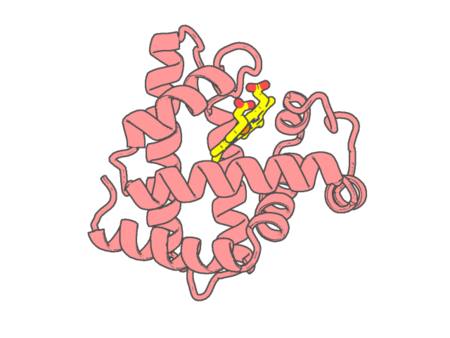 |
Experimental method | X-RAY DIFFRACTION |
|---|---|---|
| Structural data | available | |
| Non-standard amino acids | None | |
| Missing residues/ Structural gap | None / None | |
| Resolution | 2 Å | |
| Deposition date | 2009-05-09 |
| pdb_00003hep | Ferric Horse Heart Myoglobin; H64V Mutant, Nitrite Modified | ||
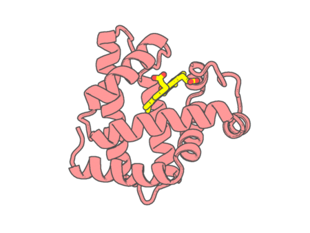 |
Experimental method | X-RAY DIFFRACTION |
|---|---|---|
| Structural data | available | |
| Non-standard amino acids | None | |
| Missing residues/ Structural gap | None / None | |
| Resolution | 1.95 Å | |
| Deposition date | 2009-05-09 |
| pdb_00003hf2 | Crystal structure of the I401P mutant of cytochrome P450 BM3 | ||
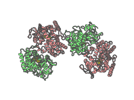 |
Experimental method | X-RAY DIFFRACTION |
|---|---|---|
| Structural data | available | |
| Non-standard amino acids | None | |
| Missing residues/ Structural gap | Exists / Exists | |
| Resolution | 2.2 Å | |
| Deposition date | 2009-05-10 |
| pdb_00003hf4 | Crystal structure of rat methemoglobin in R2 state | ||
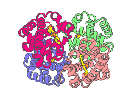 |
Experimental method | X-RAY DIFFRACTION |
|---|---|---|
| Structural data | available | |
| Non-standard amino acids | None | |
| Missing residues/ Structural gap | None / None | |
| Resolution | 2.7 Å | |
| Deposition date | 2009-05-11 |
| pdb_00003hhb | THE CRYSTAL STRUCTURE OF HUMAN DEOXYHAEMOGLOBIN AT 1.74 ANGSTROMS RESOLUTION | ||
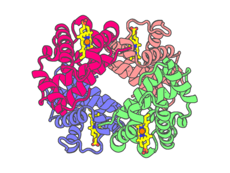 |
Experimental method | X-RAY DIFFRACTION |
|---|---|---|
| Structural data | available | |
| Non-standard amino acids | None | |
| Missing residues/ Structural gap | None / None | |
| Resolution | 1.74 Å | |
| Deposition date | 1984-03-07 |
| pdb_00003hni | Crystal structure of the Zn-induced tetramer of the engineered cyt cb562 variant RIDC-1 | ||
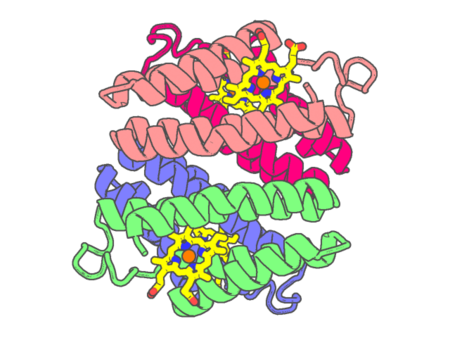 |
Experimental method | X-RAY DIFFRACTION |
|---|---|---|
| Structural data | available | |
| Non-standard amino acids | None | |
| Missing residues/ Structural gap | None / None | |
| Resolution | 2.35 Å | |
| Deposition date | 2009-05-31 |
| pdb_00003hnj | Crystal structure of the Zn-induced tetramer of the engineered cyt cb562 variant RIDC-2 | ||
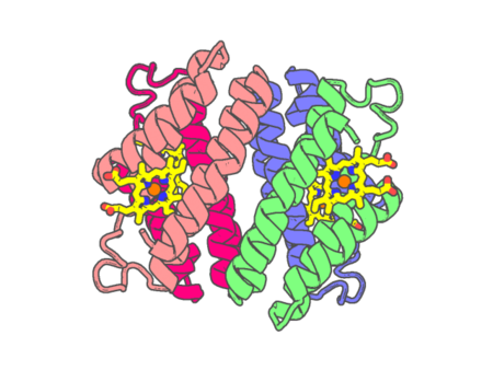 |
Experimental method | X-RAY DIFFRACTION |
|---|---|---|
| Structural data | available | |
| Non-standard amino acids | None | |
| Missing residues/ Structural gap | None / None | |
| Resolution | 2 Å | |
| Deposition date | 2009-05-31 |
| pdb_00003hnk | Crystal structure of the dimeric assembly of the cyt cb562 variant RIDC-1 | ||
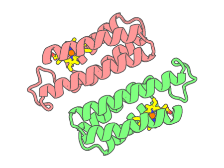 |
Experimental method | X-RAY DIFFRACTION |
|---|---|---|
| Structural data | available | |
| Non-standard amino acids | None | |
| Missing residues/ Structural gap | None / None | |
| Resolution | 2.1 Å | |
| Deposition date | 2009-05-31 |