
Number of data: 7660
Dataset: all data
| pdb_00003ml1 | Crystal Structure of the Periplasmic Nitrate Reductase from Cupriavidus necator | ||
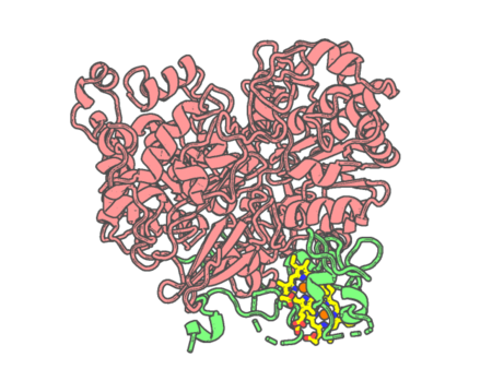 |
Experimental method | X-RAY DIFFRACTION |
|---|---|---|
| Structural data | available | |
| Non-standard amino acids | None | |
| Missing residues/ Structural gap | Exists / Exists | |
| Resolution | 1.6 Å | |
| Deposition date | 2010-04-16 |
| pdb_00003mm1 | Dye-decolorizing peroxidase (DyP) D171N | ||
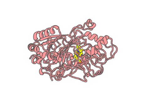 |
Experimental method | X-RAY DIFFRACTION |
|---|---|---|
| Structural data | available | |
| Non-standard amino acids | None | |
| Missing residues/ Structural gap | None / None | |
| Resolution | 1.42 Å | |
| Deposition date | 2010-04-19 |
| pdb_00003mm2 | Dye-decolorizing peroxidase (DyP) in complex with cyanide | ||
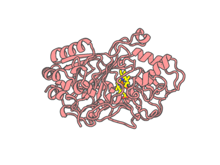 |
Experimental method | X-RAY DIFFRACTION |
|---|---|---|
| Structural data | available | |
| Non-standard amino acids | None | |
| Missing residues/ Structural gap | None / None | |
| Resolution | 1.45 Å | |
| Deposition date | 2010-04-19 |
| pdb_00003mm3 | Dye-decolorizing peroxidase (DyP) D171N in complex with cyanide | ||
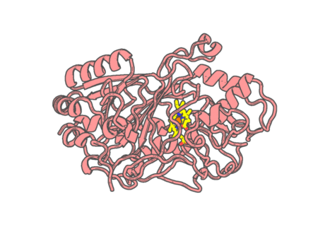 |
Experimental method | X-RAY DIFFRACTION |
|---|---|---|
| Structural data | available | |
| Non-standard amino acids | None | |
| Missing residues/ Structural gap | None / None | |
| Resolution | 1.4 Å | |
| Deposition date | 2010-04-19 |
| pdb_00003mmo | Structure of the Thioalkalivibrio nitratireducens cytochrome c nitrite reductase in complex with cyanide | ||
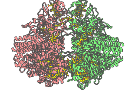 |
Experimental method | X-RAY DIFFRACTION |
|---|---|---|
| Structural data | available | |
| Non-standard amino acids | None | |
| Missing residues/ Structural gap | None / None | |
| Resolution | 1.55 Å | |
| Deposition date | 2010-04-20 |
| pdb_00003mn0 | Introducing a 2-His-1-Glu Non-Heme Iron Center into Myoglobin confers Nitric Oxide Reductase activity: Cu(II)-CN-FeBMb(-His) form | ||
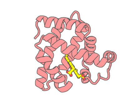 |
Experimental method | X-RAY DIFFRACTION |
|---|---|---|
| Structural data | available | |
| Non-standard amino acids | None | |
| Missing residues/ Structural gap | None / None | |
| Resolution | 1.65 Å | |
| Deposition date | 2010-04-20 |
| pdb_00003mol | Structure of dimeric holo HasAp H32A Mutant from Pseudomonas aeruginosa to 1.20A Resolution | ||
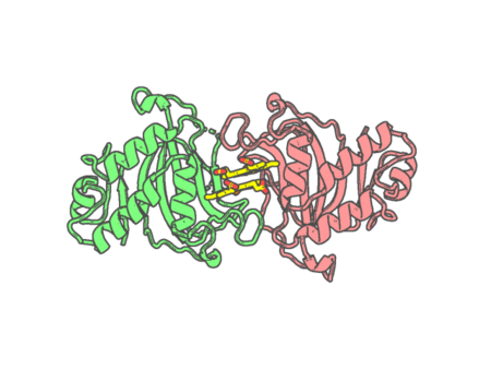 |
Experimental method | X-RAY DIFFRACTION |
|---|---|---|
| Structural data | available | |
| Non-standard amino acids | None | |
| Missing residues/ Structural gap | Exists / Exists | |
| Resolution | 1.2 Å | |
| Deposition date | 2010-04-22 |
| pdb_00003mom | Structure of holo HasAp H32A mutant complexed with imidazole from Pseudomonas aeruginosa to 2.25A Resolution | ||
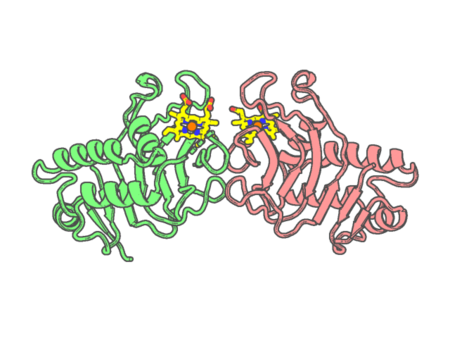 |
Experimental method | X-RAY DIFFRACTION |
|---|---|---|
| Structural data | available | |
| Non-standard amino acids | None | |
| Missing residues/ Structural gap | Exists / Exists | |
| Resolution | 2.25 Å | |
| Deposition date | 2010-04-22 |
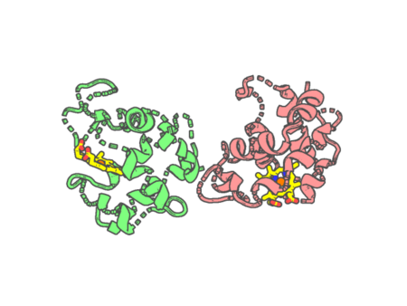
| pdb_00003mou | Characterization of the Inhibitor Binding Site of the Dehaloperoxidase-Hemoglobin from Amphitrite ornata using High-Pressure Xenon Derivatization | ||
 |
Experimental method | X-RAY DIFFRACTION |
|---|---|---|
| Structural data | available | |
| Non-standard amino acids | None | |
| Missing residues/ Structural gap | None / None | |
| Resolution | 1.711 Å | |
| Deposition date | 2010-04-23 |
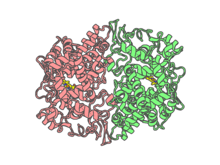
| pdb_00003mqe | Structure of SC-75416 bound at the COX-2 active site | ||
 |
Experimental method | X-RAY DIFFRACTION |
|---|---|---|
| Structural data | available | |
| Non-standard amino acids | None | |
| Missing residues/ Structural gap | None / None | |
| Resolution | 2.8 Å | |
| Deposition date | 2010-04-28 |
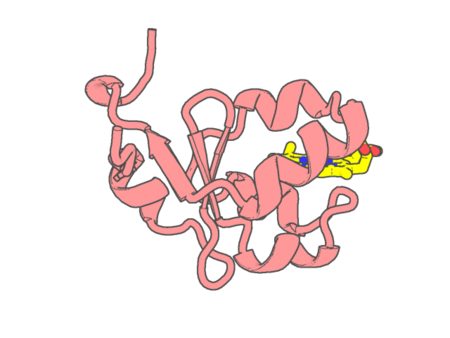
| pdb_00003mus | 2A Resolution Structure of Rat Type B Cytochrome b5 | ||
 |
Experimental method | X-RAY DIFFRACTION |
|---|---|---|
| Structural data | available | |
| Non-standard amino acids | None | |
| Missing residues/ Structural gap | None / None | |
| Resolution | 2 Å | |
| Deposition date | 2010-05-03 |
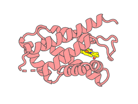
| pdb_00003mvc | High resolution crystal structure of the heme domain of GLB-6 from C. elegans | ||
 |
Experimental method | X-RAY DIFFRACTION |
|---|---|---|
| Structural data | available | |
| Non-standard amino acids | None | |
| Missing residues/ Structural gap | Exists / Exists | |
| Resolution | 1.401 Å | |
| Deposition date | 2010-05-03 |
| pdb_00003mvf | Crystal Structure of Nitrophorin 4 from Rhodnius prolixus Complexed with Nitrite at pH 7.4 | ||
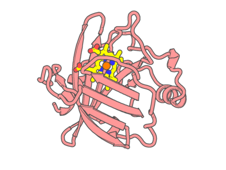 |
Experimental method | X-RAY DIFFRACTION |
|---|---|---|
| Structural data | available | |
| Non-standard amino acids | None | |
| Missing residues/ Structural gap | None / None | |
| Resolution | 1.4 Å | |
| Deposition date | 2010-05-04 |
| pdb_00003mvr | Crystal Structure of cytochrome P450 2B4-H226Y in a closed conformation | ||
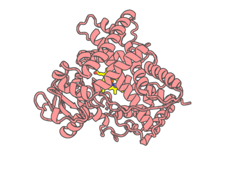 |
Experimental method | X-RAY DIFFRACTION |
|---|---|---|
| Structural data | available | |
| Non-standard amino acids | None | |
| Missing residues/ Structural gap | Exists / Exists | |
| Resolution | 1.76 Å | |
| Deposition date | 2010-05-04 |
| pdb_00003mym | Mutation of Methionine-86 in Dehaloperoxidase-hemoglobin: Effects of the Asp-His-Fe Triad in a 3/3 Globin | ||
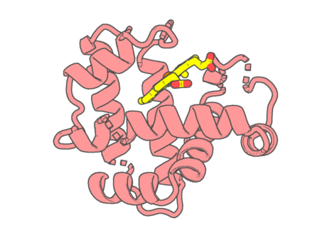 |
Experimental method | X-RAY DIFFRACTION |
|---|---|---|
| Structural data | available | |
| Non-standard amino acids | None | |
| Missing residues/ Structural gap | None / None | |
| Resolution | 1.72 Å | |
| Deposition date | 2010-05-10 |
| pdb_00003myn | Mutation of Methionine-86 in Dehaloperoxidase-hemoglobin: Effects of the Asp-His-Fe Triad in a 3/3 Globin | ||
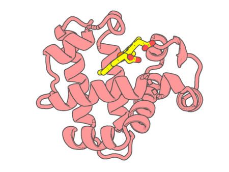 |
Experimental method | X-RAY DIFFRACTION |
|---|---|---|
| Structural data | available | |
| Non-standard amino acids | None | |
| Missing residues/ Structural gap | None / None | |
| Resolution | 2.194 Å | |
| Deposition date | 2010-05-10 |
| pdb_00003mzs | Crystal Structure of Cytochrome P450 CYP11A1 in complex with 22-hydroxy-cholesterol | ||
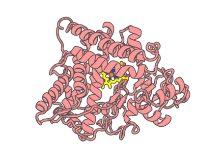 |
Experimental method | X-RAY DIFFRACTION |
|---|---|---|
| Structural data | available | |
| Non-standard amino acids | None | |
| Missing residues/ Structural gap | None / None | |
| Resolution | 2.5 Å | |
| Deposition date | 2010-05-12 |
| pdb_00003n2r | Structure of neuronal nitric oxide synthase heme domain in complex with 6-(((3R,4R/3S,4S)-4-(3-Phenoxyphenoxy)pyrrolidin-3-yl)methyl)pyridin-2-amine | ||
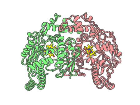 |
Experimental method | X-RAY DIFFRACTION |
|---|---|---|
| Structural data | available | |
| Non-standard amino acids | None | |
| Missing residues/ Structural gap | Exists / Exists | |
| Resolution | 1.9 Å | |
| Deposition date | 2010-05-18 |
| pdb_00003n5p | Structure of endothelial nitric oxide synthase heme domain complexed with 4-(2-(6-(2-(6-amino-4-methylpyridin-2-yl)ethyl)pyridin-2-yl)ethyl)-6-methylpyridin-2-amine | ||
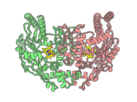 |
Experimental method | X-RAY DIFFRACTION |
|---|---|---|
| Structural data | available | |
| Non-standard amino acids | None | |
| Missing residues/ Structural gap | Exists / Exists | |
| Resolution | 2.39 Å | |
| Deposition date | 2010-05-25 |
| pdb_00003n5q | Structure of endothelial nitric oxide synthase heme domain complexed with 6,6'-(2,2'-(5-amino-1,3-phenylene)bis(ethane-2,1-diyl))bis(4-methylpyridin-2-amine) | ||
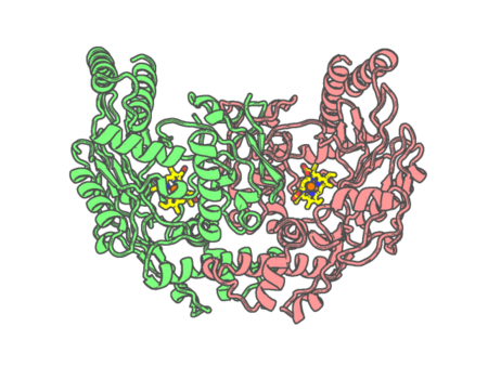 |
Experimental method | X-RAY DIFFRACTION |
|---|---|---|
| Structural data | available | |
| Non-standard amino acids | None | |
| Missing residues/ Structural gap | Exists / Exists | |
| Resolution | 2.9 Å | |
| Deposition date | 2010-05-25 |