Number of data: 3898
Dataset: (exptl_method: X-ray) and (resolution_limit=2.0)
| pdb_00006ekx |
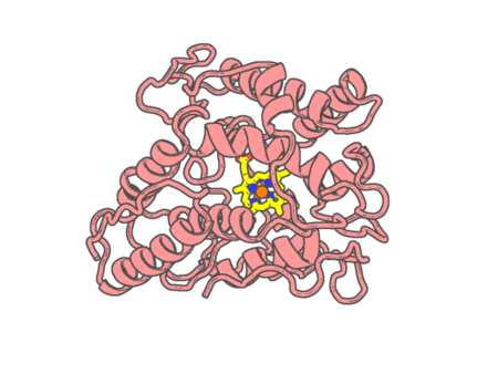
Direct-evolutioned unspecific peroxygenase from Agrocybe aegerita, in complex with 1-naphthol (I)
|
 |
Experimental method |
X-RAY DIFFRACTION |
| Structural data |
available |
| Non-standard amino acids |
None |
| Missing residues/ Structural gap |
None / None |
| Resolution |
1.13 Å |
| Deposition date |
2017-09-27 |
| pdb_00006eky |
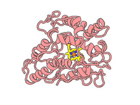
Direct-evolutioned unspecific peroxygenase from Agrocybe aegerita, in complex with 1-naphthol (II)
|
 |
Experimental method |
X-RAY DIFFRACTION |
| Structural data |
available |
| Non-standard amino acids |
None |
| Missing residues/ Structural gap |
None / None |
| Resolution |
1.23 Å |
| Deposition date |
2017-09-27 |
| pdb_00006ekz |
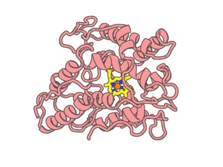
Direct-evolutioned unspecific peroxygenase from Agrocybe aegerita, in complex with propranolol
|
 |
Experimental method |
X-RAY DIFFRACTION |
| Structural data |
available |
| Non-standard amino acids |
None |
| Missing residues/ Structural gap |
None / None |
| Resolution |
1.08 Å |
| Deposition date |
2017-09-27 |
| pdb_00006el0 |
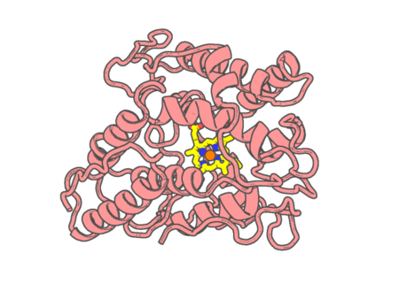
Direct-evolutioned unspecific peroxygenase from Agrocybe aegerita, in complex with styrene
|
 |
Experimental method |
X-RAY DIFFRACTION |
| Structural data |
available |
| Non-standard amino acids |
None |
| Missing residues/ Structural gap |
None / None |
| Resolution |
1.65 Å |
| Deposition date |
2017-09-27 |
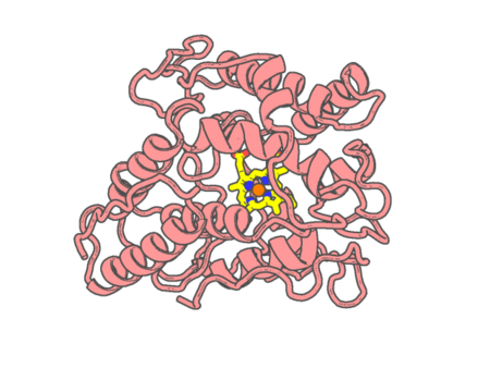
| pdb_00006el4 |
Direct-evolutioned unspecific peroxygenase from Agrocybe aegerita, in complex with veratryl alcohol
|
 |
Experimental method |
X-RAY DIFFRACTION |
| Structural data |
available |
| Non-standard amino acids |
None |
| Missing residues/ Structural gap |
None / None |
| Resolution |
1.53 Å |
| Deposition date |
2017-09-27 |
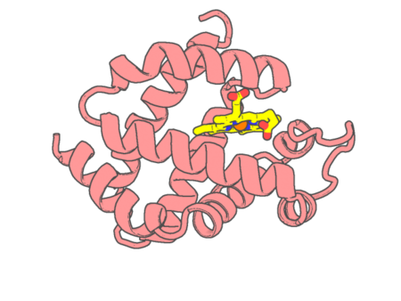
| pdb_00006eye |
Crystal structure of murine neuroglobin under 150 bar krypton
|
 |
Experimental method |
X-RAY DIFFRACTION |
| Structural data |
available |
| Non-standard amino acids |
None |
| Missing residues/ Structural gap |
None / None |
| Resolution |
1.7 Å |
| Deposition date |
2017-11-12 |
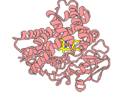
| pdb_00006f0c |
Cytochrome P450 TxtC employs substrate conformational switching for sequential aliphatic and aromatic thaxtomin hydroxylation
|
 |
Experimental method |
X-RAY DIFFRACTION |
| Structural data |
available |
| Non-standard amino acids |
None |
| Missing residues/ Structural gap |
None / None |
| Resolution |
1.7 Å |
| Deposition date |
2017-11-17 |
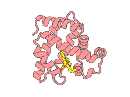
| pdb_00006f17 |
Structure of Mb NMH H64V, V68A mutant resting state
|
 |
Experimental method |
X-RAY DIFFRACTION |
| Structural data |
available |
| Non-standard amino acids |
Included |
| Missing residues/ Structural gap |
None / None |
| Resolution |
1.45 Å |
| Deposition date |
2017-11-21 |
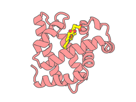
| pdb_00006f18 |
Structure of Mb NMH H64V, V68A mutant complex with EDA
|
 |
Experimental method |
X-RAY DIFFRACTION |
| Structural data |
available |
| Non-standard amino acids |
Included |
| Missing residues/ Structural gap |
None / None |
| Resolution |
1.8 Å |
| Deposition date |
2017-11-21 |
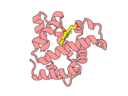
| pdb_00006f19 |
Structure of Mb NMH H64V, V68A mutant complex with EDA incubated at room temperature for 5 min
|
 |
Experimental method |
X-RAY DIFFRACTION |
| Structural data |
available |
| Non-standard amino acids |
Included |
| Missing residues/ Structural gap |
None / None |
| Resolution |
1.895 Å |
| Deposition date |
2017-11-21 |
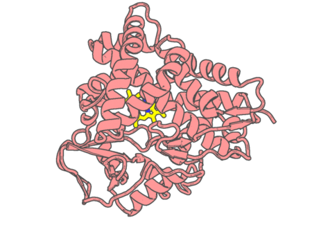
| pdb_00006f88 |
Crystal structure of cytochrome P450 CYP260A1 (S276N) bound with progesterone
|
 |
Experimental method |
X-RAY DIFFRACTION |
| Structural data |
available |
| Non-standard amino acids |
None |
| Missing residues/ Structural gap |
None / None |
| Resolution |
1.75 Å |
| Deposition date |
2017-12-12 |
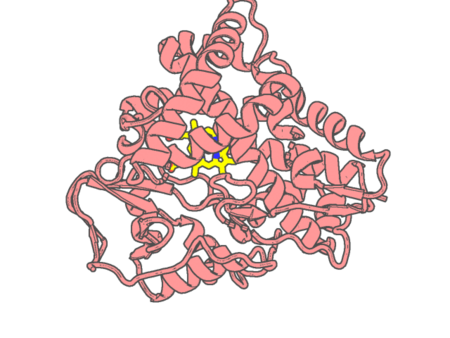
| pdb_00006f8a |
Crystal structure of cytochrome P450 CYP260A1 (S276I) bound with histidine
|
 |
Experimental method |
X-RAY DIFFRACTION |
| Structural data |
available |
| Non-standard amino acids |
None |
| Missing residues/ Structural gap |
None / None |
| Resolution |
1.35 Å |
| Deposition date |
2017-12-12 |
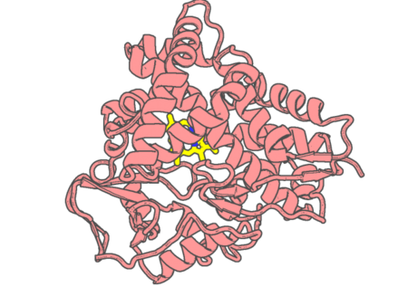
| pdb_00006f8c |
Crystal structure of cytochrome P450 CYP260A1 (S276I) bound with progesterone
|
 |
Experimental method |
X-RAY DIFFRACTION |
| Structural data |
available |
| Non-standard amino acids |
None |
| Missing residues/ Structural gap |
None / None |
| Resolution |
1.9 Å |
| Deposition date |
2017-12-12 |
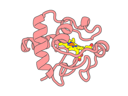
| pdb_00006ff5 |
X-ray structure of bovine heart cytochrome c at high ionic strength
|
 |
Experimental method |
X-RAY DIFFRACTION |
| Structural data |
available |
| Non-standard amino acids |
None |
| Missing residues/ Structural gap |
None / None |
| Resolution |
1.74 Å |
| Deposition date |
2018-01-03 |
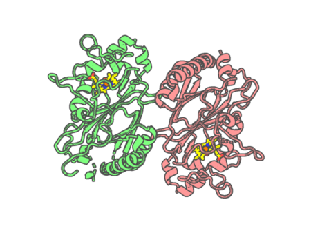
| pdb_00006fiy |
Crystal structure of a dye-decolorizing peroxidase D143AR232A variant from Klebsiella pneumoniae (KpDyP)
|
 |
Experimental method |
X-RAY DIFFRACTION |
| Structural data |
available |
| Non-standard amino acids |
None |
| Missing residues/ Structural gap |
None / None |
| Resolution |
1.09000433235 Å |
| Deposition date |
2018-01-19 |
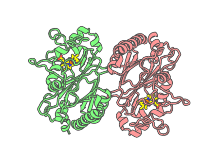
| pdb_00006fks |
Crystal structure of a dye-decolorizing peroxidase from Klebsiella pneumoniae (KpDyP)
|
 |
Experimental method |
X-RAY DIFFRACTION |
| Structural data |
available |
| Non-standard amino acids |
None |
| Missing residues/ Structural gap |
None / None |
| Resolution |
1.60000463928 Å |
| Deposition date |
2018-01-24 |
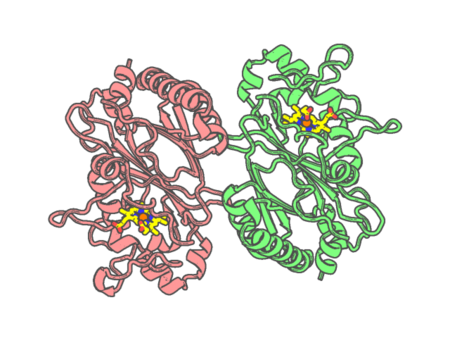
| pdb_00006fkt |
Crystal structure of a dye-decolorizing peroxidase R232A variant from Klebsiella pneumoniae (KpDyP)
|
 |
Experimental method |
X-RAY DIFFRACTION |
| Structural data |
available |
| Non-standard amino acids |
None |
| Missing residues/ Structural gap |
None / None |
| Resolution |
1.86002782211 Å |
| Deposition date |
2018-01-24 |
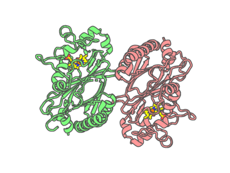
| pdb_00006fl2 |
Crystal structure of a dye-decolorizing peroxidase D143A variant from Klebsiella pneumoniae (KpDyP)
|
 |
Experimental method |
X-RAY DIFFRACTION |
| Structural data |
available |
| Non-standard amino acids |
None |
| Missing residues/ Structural gap |
None / None |
| Resolution |
1.27000110594 Å |
| Deposition date |
2018-01-25 |
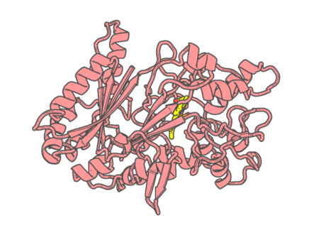
| pdb_00006fsk |
F194Y mutant of the Dye-decolorizing peroxidase (DYP) from Pleurotus ostreatus
|
 |
Experimental method |
X-RAY DIFFRACTION |
| Structural data |
available |
| Non-standard amino acids |
Included |
| Missing residues/ Structural gap |
None / None |
| Resolution |
1.56 Å |
| Deposition date |
2018-02-19 |
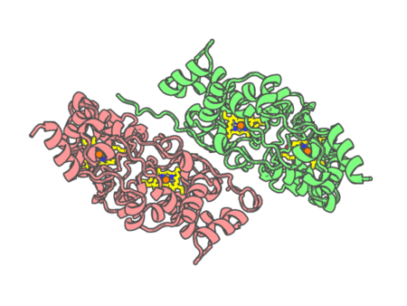
| pdb_00006fu3 |
Structure of the mixed-valence, active form, of cytochrome c peroxidase from obligate human pathogenic bacterium Neisseria gonorrhoeae
|
 |
Experimental method |
X-RAY DIFFRACTION |
| Structural data |
available |
| Non-standard amino acids |
None |
| Missing residues/ Structural gap |
None / None |
| Resolution |
1.8 Å |
| Deposition date |
2018-02-26 |
