
Number of data: 3898
Dataset: (exptl_method: X-ray) and (resolution_limit=2.0)
| pdb_00006h6i | Ferric murine neuroglobin Gly-loop mutant | ||
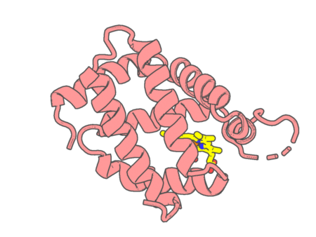 |
Experimental method | X-RAY DIFFRACTION |
|---|---|---|
| Structural data | available | |
| Non-standard amino acids | None | |
| Missing residues/ Structural gap | Exists / Exists | |
| Resolution | 1.6 Å | |
| Deposition date | 2018-07-27 |
| pdb_00006hbi | SCAPHARCA DIMERIC HEMOGLOBIN, MUTANT T72V, DEOXY FORM | ||
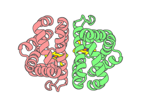 |
Experimental method | X-RAY DIFFRACTION |
|---|---|---|
| Structural data | available | |
| Non-standard amino acids | None | |
| Missing residues/ Structural gap | None / None | |
| Resolution | 1.8 Å | |
| Deposition date | 1998-06-25 |
| pdb_00006hbw | Crystal structure of deoxy-human hemoglobin beta6 glu->trp | ||
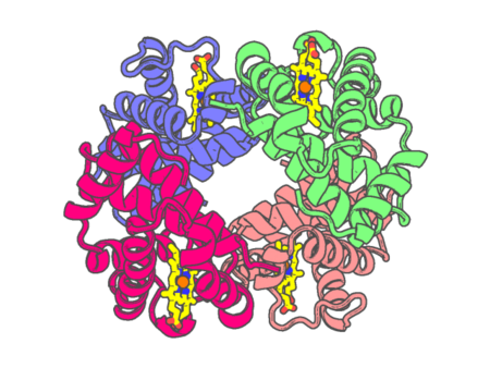 |
Experimental method | X-RAY DIFFRACTION |
|---|---|---|
| Structural data | available | |
| Non-standard amino acids | None | |
| Missing residues/ Structural gap | None / None | |
| Resolution | 2 Å | |
| Deposition date | 1998-09-10 |
| pdb_00006hih | Cytochrome c prime beta from Methylococcus capsulatus (Bath) | ||
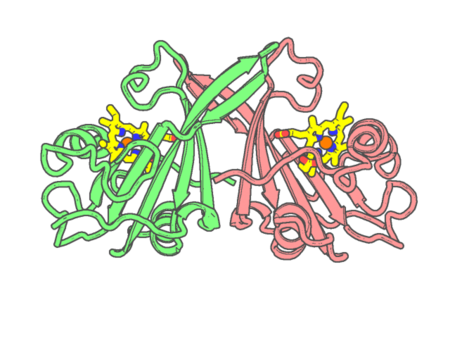 |
Experimental method | X-RAY DIFFRACTION |
|---|---|---|
| Structural data | available | |
| Non-standard amino acids | None | |
| Missing residues/ Structural gap | None / None | |
| Resolution | 1.6 Å | |
| Deposition date | 2018-08-29 |
| pdb_00006hiu | Cytochrome P460 from Methylococcus capsulatus (Bath) | ||
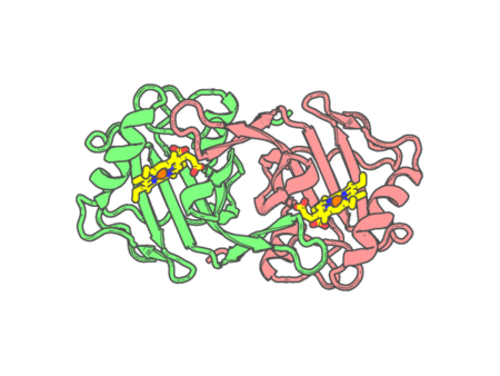 |
Experimental method | X-RAY DIFFRACTION |
|---|---|---|
| Structural data | available | |
| Non-standard amino acids | None | |
| Missing residues/ Structural gap | None / None | |
| Resolution | 1.36 Å | |
| Deposition date | 2018-08-31 |
| pdb_00006hk2 | Crystal structure of ferric R-state human methemoglobin bound to maleimide-deferoxamine bifunctional chelator (DFO) | ||
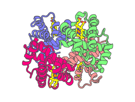 |
Experimental method | X-RAY DIFFRACTION |
|---|---|---|
| Structural data | available | |
| Non-standard amino acids | None | |
| Missing residues/ Structural gap | None / None | |
| Resolution | 1.55 Å | |
| Deposition date | 2018-09-05 |
| pdb_00006hn8 | Structure of BM3 heme domain in complex with troglitazone | ||
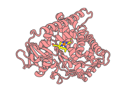 |
Experimental method | X-RAY DIFFRACTION |
|---|---|---|
| Structural data | available | |
| Non-standard amino acids | None | |
| Missing residues/ Structural gap | None / None | |
| Resolution | 1.997 Å | |
| Deposition date | 2018-09-14 |
| pdb_00006hqd | Cytochrome P450-153 from Pseudomonas sp. 19-rlim | ||
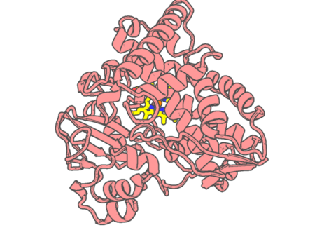 |
Experimental method | X-RAY DIFFRACTION |
|---|---|---|
| Structural data | available | |
| Non-standard amino acids | None | |
| Missing residues/ Structural gap | None / None | |
| Resolution | 1.8 Å | |
| Deposition date | 2018-09-24 |
| pdb_00006hqk | Crystal structure of GcoA F169A bound to guaiacol | ||
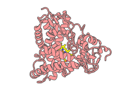 |
Experimental method | X-RAY DIFFRACTION |
|---|---|---|
| Structural data | available | |
| Non-standard amino acids | None | |
| Missing residues/ Structural gap | None / None | |
| Resolution | 1.57 Å | |
| Deposition date | 2018-09-25 |
| pdb_00006hql | Crystal structure of GcoA F169H bound to guaiacol | ||
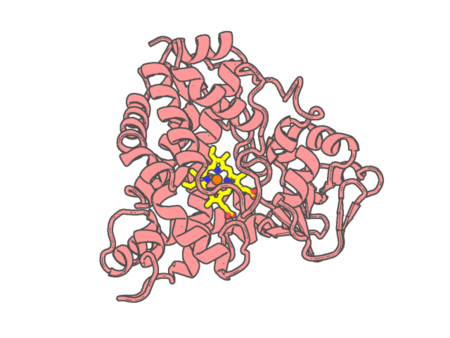 |
Experimental method | X-RAY DIFFRACTION |
|---|---|---|
| Structural data | available | |
| Non-standard amino acids | None | |
| Missing residues/ Structural gap | None / None | |
| Resolution | 1.49 Å | |
| Deposition date | 2018-09-25 |
| pdb_00006hqm | Crystal structure of GcoA F169I bound to guaiacol | ||
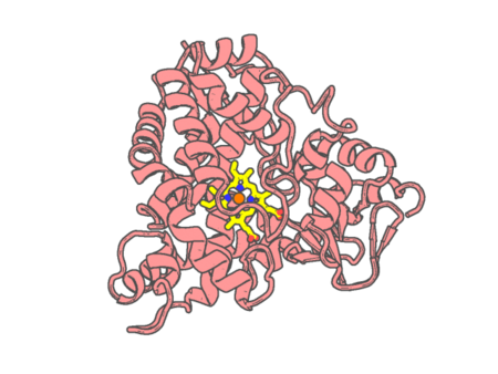 |
Experimental method | X-RAY DIFFRACTION |
|---|---|---|
| Structural data | available | |
| Non-standard amino acids | None | |
| Missing residues/ Structural gap | None / None | |
| Resolution | 1.85 Å | |
| Deposition date | 2018-09-25 |
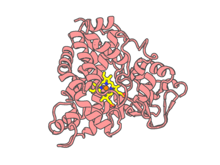
| pdb_00006hqn | Crystal structure of GcoA F169L bound to guaiacol | ||
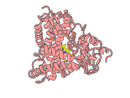 |
Experimental method | X-RAY DIFFRACTION |
|---|---|---|
| Structural data | available | |
| Non-standard amino acids | None | |
| Missing residues/ Structural gap | None / None | |
| Resolution | 1.87 Å | |
| Deposition date | 2018-09-25 |
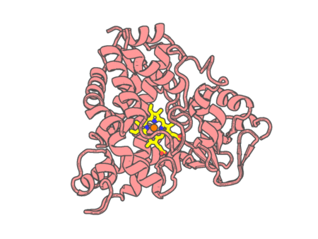
| pdb_00006hqo | Crystal structure of GcoA F169S bound to guaiacol | ||
 |
Experimental method | X-RAY DIFFRACTION |
|---|---|---|
| Structural data | available | |
| Non-standard amino acids | None | |
| Missing residues/ Structural gap | None / None | |
| Resolution | 1.7 Å | |
| Deposition date | 2018-09-25 |
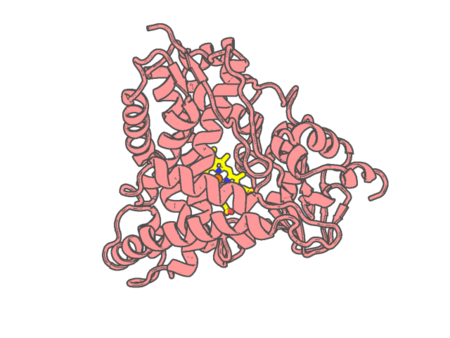
| pdb_00006hqp | Crystal structure of GcoA F169V bound to guaiacol | ||
 |
Experimental method | X-RAY DIFFRACTION |
|---|---|---|
| Structural data | available | |
| Non-standard amino acids | None | |
| Missing residues/ Structural gap | None / None | |
| Resolution | 1.62 Å | |
| Deposition date | 2018-09-25 |
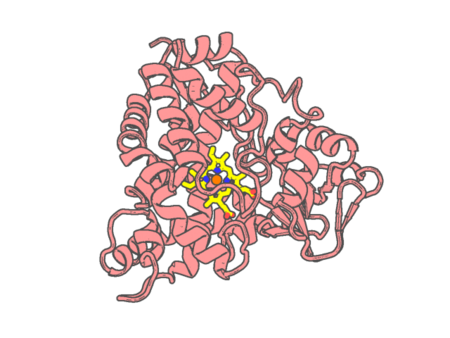
| pdb_00006hqq | Crystal structure of GcoA F169A bound to syringol | ||
 |
Experimental method | X-RAY DIFFRACTION |
|---|---|---|
| Structural data | available | |
| Non-standard amino acids | None | |
| Missing residues/ Structural gap | None / None | |
| Resolution | 1.66 Å | |
| Deposition date | 2018-09-25 |
| pdb_00006hqr | Crystal structure of GcoA F169H bound to syringol | ||
 |
Experimental method | X-RAY DIFFRACTION |
|---|---|---|
| Structural data | available | |
| Non-standard amino acids | None | |
| Missing residues/ Structural gap | None / None | |
| Resolution | 1.79 Å | |
| Deposition date | 2018-09-25 |
| pdb_00006hqt | Crystal structure of GcoA F169V bound to syringol | ||
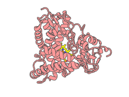 |
Experimental method | X-RAY DIFFRACTION |
|---|---|---|
| Structural data | available | |
| Non-standard amino acids | None | |
| Missing residues/ Structural gap | None / None | |
| Resolution | 1.85 Å | |
| Deposition date | 2018-09-25 |
| pdb_00006hr0 | Optimizing electroactive organisms: the effect of orthologous proteins | ||
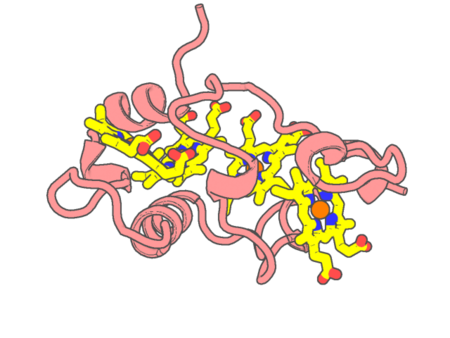 |
Experimental method | X-RAY DIFFRACTION |
|---|---|---|
| Structural data | available | |
| Non-standard amino acids | None | |
| Missing residues/ Structural gap | None / None | |
| Resolution | 1.04 Å | |
| Deposition date | 2018-09-26 |
| pdb_00006i3t | Crystal structure of murine neuroglobin bound to CO at 40 K. | ||
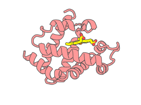 |
Experimental method | X-RAY DIFFRACTION |
|---|---|---|
| Structural data | available | |
| Non-standard amino acids | None | |
| Missing residues/ Structural gap | None / None | |
| Resolution | 2 Å | |
| Deposition date | 2018-11-07 |
| pdb_00006i40 | Crystal structure of murine neuroglobin bound to CO at 15K under illumination using optical fiber | ||
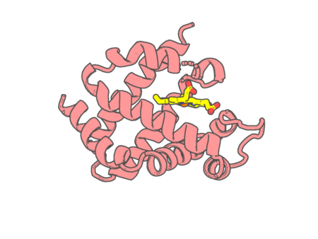 |
Experimental method | X-RAY DIFFRACTION |
|---|---|---|
| Structural data | available | |
| Non-standard amino acids | None | |
| Missing residues/ Structural gap | None / None | |
| Resolution | 1.9 Å | |
| Deposition date | 2018-11-08 |