
Number of data: 7660
Dataset: all data
| pdb_00003nod | MURINE INDUCIBLE NITRIC OXIDE SYNTHASE OXYGENASE DIMER (DELTA 65) WITH TETRAHYDROBIOPTERIN AND PRODUCT ANALOGUE L-THIOCITRULLINE | ||
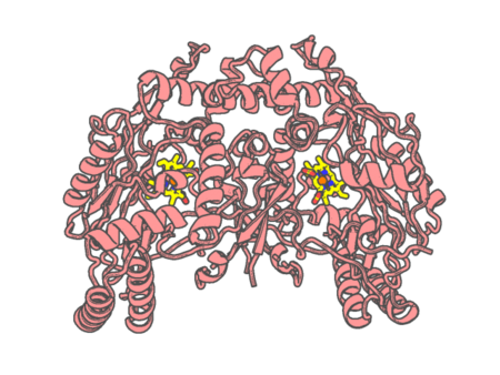 |
Experimental method | X-RAY DIFFRACTION |
|---|---|---|
| Structural data | available | |
| Non-standard amino acids | None | |
| Missing residues/ Structural gap | Exists / Exists | |
| Resolution | 2.7 Å | |
| Deposition date | 1998-03-06 |
| pdb_00003nos | HUMAN ENDOTHELIAL NITRIC OXIDE SYNTHASE WITH ARGININE SUBSTRATE | ||
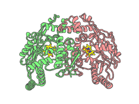 |
Experimental method | X-RAY DIFFRACTION |
|---|---|---|
| Structural data | available | |
| Non-standard amino acids | None | |
| Missing residues/ Structural gap | Exists / Exists | |
| Resolution | 2.4 Å | |
| Deposition date | 1999-02-03 |
| pdb_00003np1 | CRYSTAL STRUCTURE OF THE COMPLEX OF NITROPHORIN 1 FROM RHODNIUS PROLIXUS WITH CYANIDE | ||
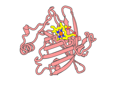 |
Experimental method | X-RAY DIFFRACTION |
|---|---|---|
| Structural data | available | |
| Non-standard amino acids | None | |
| Missing residues/ Structural gap | None / None | |
| Resolution | 2.3 Å | |
| Deposition date | 1998-01-22 |
| pdb_00003npl | Structure of Ru(bpy)2(A-Phen)(K97C) P450 BM3 heme domain, a ruthenium modified P450 BM3 mutant | ||
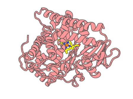 |
Experimental method | X-RAY DIFFRACTION |
|---|---|---|
| Structural data | available | |
| Non-standard amino acids | None | |
| Missing residues/ Structural gap | None / None | |
| Resolution | 2.4 Å | |
| Deposition date | 2010-06-28 |
| pdb_00003nqs | Crystal Structure of Inducible Nitric Oxide Synthase with N-Nitrosated-pterin | ||
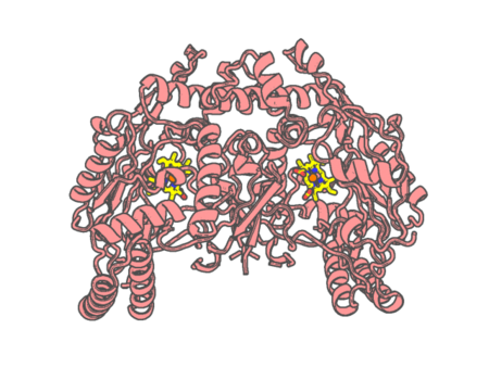 |
Experimental method | X-RAY DIFFRACTION |
|---|---|---|
| Structural data | available | |
| Non-standard amino acids | None | |
| Missing residues/ Structural gap | Exists / Exists | |
| Resolution | 2.2 Å | |
| Deposition date | 2010-06-29 |
| pdb_00003nse | BOVINE ENOS, H4B-FREE, SEITU COMPLEX | ||
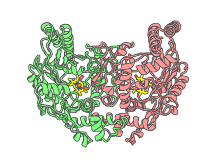 |
Experimental method | X-RAY DIFFRACTION |
|---|---|---|
| Structural data | available | |
| Non-standard amino acids | None | |
| Missing residues/ Structural gap | None / None | |
| Resolution | 2.1 Å | |
| Deposition date | 1998-09-23 |
| pdb_00003nt1 | High resolution structure of naproxen:COX-2 complex. | ||
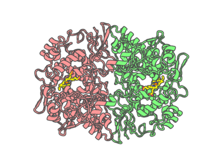 |
Experimental method | X-RAY DIFFRACTION |
|---|---|---|
| Structural data | available | |
| Non-standard amino acids | None | |
| Missing residues/ Structural gap | None / None | |
| Resolution | 1.73 Å | |
| Deposition date | 2010-07-02 |
| pdb_00003ntb | Structure of 6-methylthio naproxen analog bound to mCOX-2. | ||
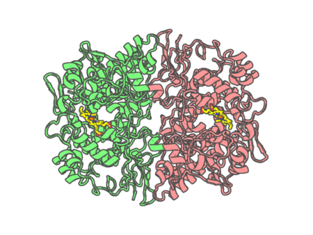 |
Experimental method | X-RAY DIFFRACTION |
|---|---|---|
| Structural data | available | |
| Non-standard amino acids | None | |
| Missing residues/ Structural gap | None / None | |
| Resolution | 2.27 Å | |
| Deposition date | 2010-07-03 |
| pdb_00003ntg | Crystal structure of COX-2 with selective compound 23d-(R) | ||
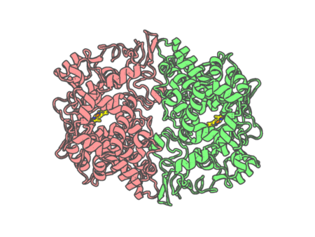 |
Experimental method | X-RAY DIFFRACTION |
|---|---|---|
| Structural data | available | |
| Non-standard amino acids | None | |
| Missing residues/ Structural gap | None / None | |
| Resolution | 2.19 Å | |
| Deposition date | 2010-07-04 |
| pdb_00003nu1 | Structure of holo form of a periplasmic heme binding protein | ||
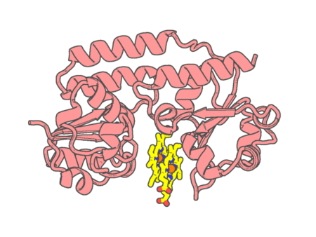 |
Experimental method | X-RAY DIFFRACTION |
|---|---|---|
| Structural data | available | |
| Non-standard amino acids | None | |
| Missing residues/ Structural gap | None / None | |
| Resolution | 2.5 Å | |
| Deposition date | 2010-07-06 |
| pdb_00003nv5 | Crystal Structure of Cytochrome P450 CYP101D2 | ||
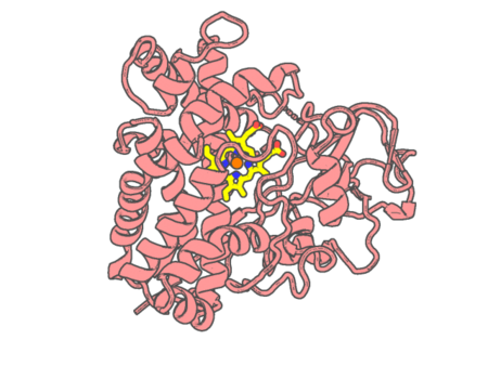 |
Experimental method | X-RAY DIFFRACTION |
|---|---|---|
| Structural data | available | |
| Non-standard amino acids | None | |
| Missing residues/ Structural gap | Exists / Exists | |
| Resolution | 2.41 Å | |
| Deposition date | 2010-07-08 |
| pdb_00003nv6 | Crystal Structure of Camphor-Bound CYP101D2 | ||
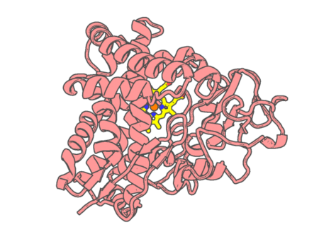 |
Experimental method | X-RAY DIFFRACTION |
|---|---|---|
| Structural data | available | |
| Non-standard amino acids | None | |
| Missing residues/ Structural gap | None / None | |
| Resolution | 2.2 Å | |
| Deposition date | 2010-07-08 |
| pdb_00003nvr | Modulating Heme Redox Potential Through Protein-Induced Porphyrin Distortion | ||
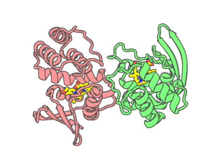 |
Experimental method | X-RAY DIFFRACTION |
|---|---|---|
| Structural data | available | |
| Non-standard amino acids | None | |
| Missing residues/ Structural gap | None / None | |
| Resolution | 2.148 Å | |
| Deposition date | 2010-07-08 |
| pdb_00003nvu | Modulating Heme Redox Potential Through Protein-Induced Porphyrin Distortion | ||
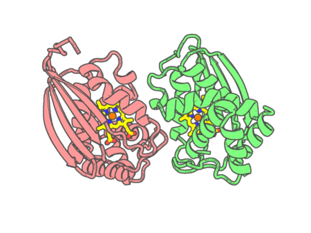 |
Experimental method | X-RAY DIFFRACTION |
|---|---|---|
| Structural data | available | |
| Non-standard amino acids | None | |
| Missing residues/ Structural gap | None / None | |
| Resolution | 2.038 Å | |
| Deposition date | 2010-07-08 |
| pdb_00003nw2 | Novel nanomolar Imidazopyridines as selective Nitric Oxide Synthase (iNOS) inhibitors: SAR and structural insights | ||
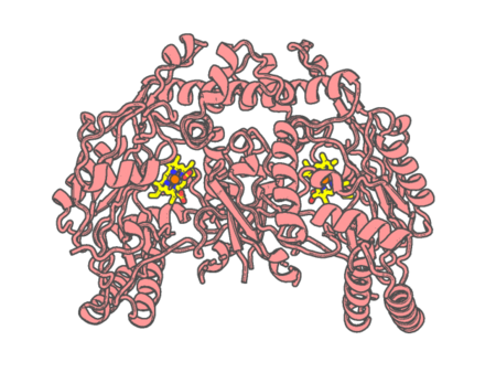 |
Experimental method | X-RAY DIFFRACTION |
|---|---|---|
| Structural data | available | |
| Non-standard amino acids | None | |
| Missing residues/ Structural gap | Exists / Exists | |
| Resolution | 2.8 Å | |
| Deposition date | 2010-07-09 |
| pdb_00003nwl | The crystal structure of the P212121 form of bovine liver catalase previously characterized by electron microscopy | ||
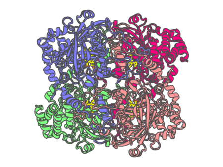 |
Experimental method | X-RAY DIFFRACTION |
|---|---|---|
| Structural data | available | |
| Non-standard amino acids | None | |
| Missing residues/ Structural gap | None / None | |
| Resolution | 2.69 Å | |
| Deposition date | 2010-07-09 |
| pdb_00003nwv | Human cytochrome c G41S | ||
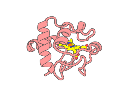 |
Experimental method | X-RAY DIFFRACTION |
|---|---|---|
| Structural data | available | |
| Non-standard amino acids | None | |
| Missing residues/ Structural gap | None / None | |
| Resolution | 1.9 Å | |
| Deposition date | 2010-07-11 |
| pdb_00003nxu | Crystal structure of human cytochrome P4503A4 bound to an inhibitor ritonavir | ||
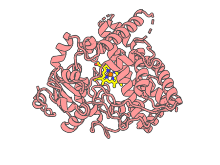 |
Experimental method | X-RAY DIFFRACTION |
|---|---|---|
| Structural data | available | |
| Non-standard amino acids | None | |
| Missing residues/ Structural gap | Exists / Exists | |
| Resolution | 2 Å | |
| Deposition date | 2010-07-14 |
| pdb_00003o0r | Crystal structure of nitric oxide reductase from Pseudomonas aeruginosa in complex with antibody fragment | ||
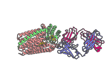 |
Experimental method | X-RAY DIFFRACTION |
|---|---|---|
| Structural data | available | |
| Non-standard amino acids | None | |
| Missing residues/ Structural gap | None / None | |
| Resolution | 2.7 Å | |
| Deposition date | 2010-07-20 |
| pdb_00003o1a | Structure of OxyE (CYP165D3), a Cytochrome P450 Involved in Teicoplanin Biosynthesis | ||
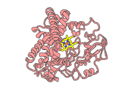 |
Experimental method | X-RAY DIFFRACTION |
|---|---|---|
| Structural data | available | |
| Non-standard amino acids | None | |
| Missing residues/ Structural gap | None / None | |
| Resolution | 2.5 Å | |
| Deposition date | 2010-07-21 |