
Number of data: 7660
Dataset: all data
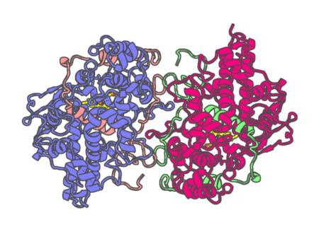
| pdb_00004dl1 | Crystal Structure of human Myeloperoxidase with covalent thioxanthine analog | ||
 |
Experimental method | X-RAY DIFFRACTION |
|---|---|---|
| Structural data | available | |
| Non-standard amino acids | Included | |
| Missing residues/ Structural gap | None / None | |
| Resolution | 2 Å | |
| Deposition date | 2012-02-05 |
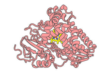
| pdb_00004dnj | The crystal structures of 4-methoxybenzoate bound CYP199A2 | ||
 |
Experimental method | X-RAY DIFFRACTION |
|---|---|---|
| Structural data | available | |
| Non-standard amino acids | None | |
| Missing residues/ Structural gap | None / None | |
| Resolution | 1.8 Å | |
| Deposition date | 2012-02-08 |
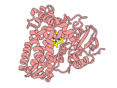
| pdb_00004dnz | The crystal structures of CYP199A4 | ||
 |
Experimental method | X-RAY DIFFRACTION |
|---|---|---|
| Structural data | available | |
| Non-standard amino acids | None | |
| Missing residues/ Structural gap | None / None | |
| Resolution | 2.6 Å | |
| Deposition date | 2012-02-09 |
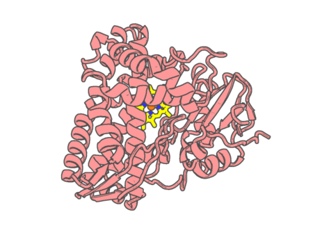
| pdb_00004do1 | The crystal structures of 4-methoxybenzoate bound CYP199A4 | ||
 |
Experimental method | X-RAY DIFFRACTION |
|---|---|---|
| Structural data | available | |
| Non-standard amino acids | None | |
| Missing residues/ Structural gap | None / None | |
| Resolution | 2 Å | |
| Deposition date | 2012-02-09 |
| pdb_00004dtw | cytochrome P450 BM3h-8C8 MRI sensor bound to serotonin | ||
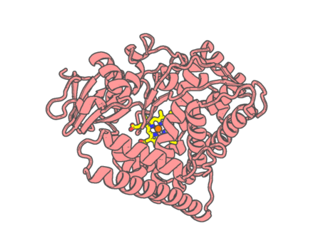 |
Experimental method | X-RAY DIFFRACTION |
|---|---|---|
| Structural data | available | |
| Non-standard amino acids | None | |
| Missing residues/ Structural gap | None / None | |
| Resolution | 1.8 Å | |
| Deposition date | 2012-02-21 |
| pdb_00004dty | cytochrome P450 BM3h-8C8 MRI sensor, no ligand | ||
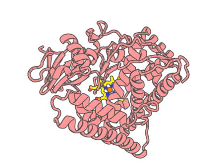 |
Experimental method | X-RAY DIFFRACTION |
|---|---|---|
| Structural data | available | |
| Non-standard amino acids | None | |
| Missing residues/ Structural gap | None / None | |
| Resolution | 1.45 Å | |
| Deposition date | 2012-02-21 |
| pdb_00004dtz | cytochrome P450 BM3h-8C8 MRI sensor bound to dopamine | ||
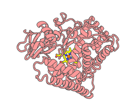 |
Experimental method | X-RAY DIFFRACTION |
|---|---|---|
| Structural data | available | |
| Non-standard amino acids | None | |
| Missing residues/ Structural gap | None / None | |
| Resolution | 1.55 Å | |
| Deposition date | 2012-02-21 |
| pdb_00004du2 | cytochrome P450 BM3h-B7 MRI sensor bound to dopamine | ||
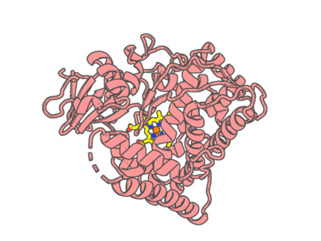 |
Experimental method | X-RAY DIFFRACTION |
|---|---|---|
| Structural data | available | |
| Non-standard amino acids | None | |
| Missing residues/ Structural gap | Exists / Exists | |
| Resolution | 1.9 Å | |
| Deposition date | 2012-02-21 |
| pdb_00004dua | cytochrome P450 BM3h-9D7 MRI sensor, no ligand | ||
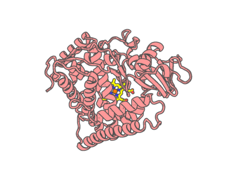 |
Experimental method | X-RAY DIFFRACTION |
|---|---|---|
| Structural data | available | |
| Non-standard amino acids | None | |
| Missing residues/ Structural gap | None / None | |
| Resolution | 2 Å | |
| Deposition date | 2012-02-21 |
| pdb_00004dub | cytochrome P450 BM3h-9D7 MRI sensor bound to dopamine | ||
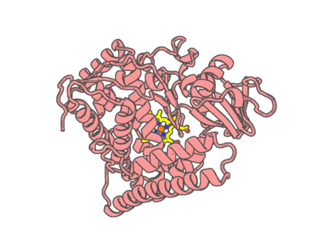 |
Experimental method | X-RAY DIFFRACTION |
|---|---|---|
| Structural data | available | |
| Non-standard amino acids | None | |
| Missing residues/ Structural gap | None / None | |
| Resolution | 1.7 Å | |
| Deposition date | 2012-02-21 |
| pdb_00004duc | cytochrome P450 BM3h-2G9 MRI sensor, no ligand | ||
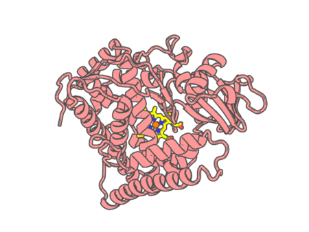 |
Experimental method | X-RAY DIFFRACTION |
|---|---|---|
| Structural data | available | |
| Non-standard amino acids | None | |
| Missing residues/ Structural gap | None / None | |
| Resolution | 1.92 Å | |
| Deposition date | 2012-02-21 |
| pdb_00004dud | cytochrome P450 BM3h-2G9C6 MRI sensor, no ligand | ||
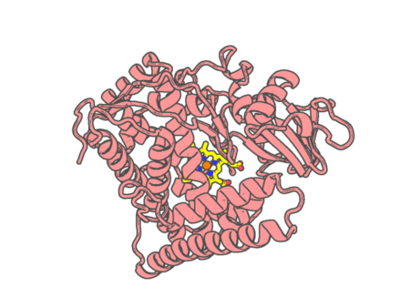 |
Experimental method | X-RAY DIFFRACTION |
|---|---|---|
| Structural data | available | |
| Non-standard amino acids | None | |
| Missing residues/ Structural gap | None / None | |
| Resolution | 1.85 Å | |
| Deposition date | 2012-02-21 |
| pdb_00004due | cytochrome P450 BM3h-2G9C6 MRI sensor bound to serotonin | ||
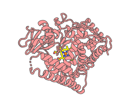 |
Experimental method | X-RAY DIFFRACTION |
|---|---|---|
| Structural data | available | |
| Non-standard amino acids | None | |
| Missing residues/ Structural gap | Exists / Exists | |
| Resolution | 1.7 Å | |
| Deposition date | 2012-02-21 |
| pdb_00004duf | cytochrome P450 BM3h-2G9 MRI sensor bound to serotonin | ||
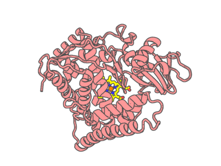 |
Experimental method | X-RAY DIFFRACTION |
|---|---|---|
| Structural data | available | |
| Non-standard amino acids | None | |
| Missing residues/ Structural gap | None / None | |
| Resolution | 1.8 Å | |
| Deposition date | 2012-02-21 |
| pdb_00004dvq | Structure of human aldosterone synthase, CYP11B2, in complex with deoxycorticosterone | ||
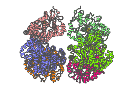 |
Experimental method | X-RAY DIFFRACTION |
|---|---|---|
| Structural data | available | |
| Non-standard amino acids | None | |
| Missing residues/ Structural gap | Exists / Exists | |
| Resolution | 2.49 Å | |
| Deposition date | 2012-02-23 |
| pdb_00004dwt | Carbonmonoxy dehaloperoxidase-hemoglobin A structure at 2.05 Angstrom resolution | ||
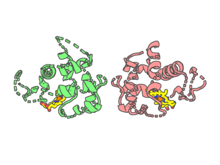 |
Experimental method | X-RAY DIFFRACTION |
|---|---|---|
| Structural data | available | |
| Non-standard amino acids | None | |
| Missing residues/ Structural gap | None / None | |
| Resolution | 2.05 Å | |
| Deposition date | 2012-02-26 |
| pdb_00004dwu | Carbonmonoxy dehaloperoxidase-hemoglobin A structure at 1.44 Angstrom resolution | ||
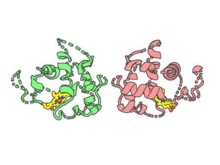 |
Experimental method | X-RAY DIFFRACTION |
|---|---|---|
| Structural data | available | |
| Non-standard amino acids | None | |
| Missing residues/ Structural gap | None / None | |
| Resolution | 1.44 Å | |
| Deposition date | 2012-02-26 |
| pdb_00004dxy | Crystal structures of CYP101D2 Y96A mutant | ||
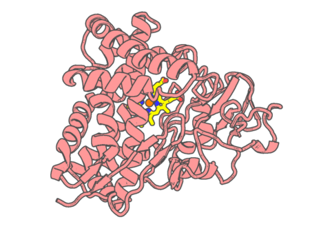 |
Experimental method | X-RAY DIFFRACTION |
|---|---|---|
| Structural data | available | |
| Non-standard amino acids | None | |
| Missing residues/ Structural gap | None / None | |
| Resolution | 2 Å | |
| Deposition date | 2012-02-28 |
| pdb_00004dy9 | Leishmania major Peroxidase is a Cytochrome c Peroxidase | ||
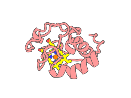 |
Experimental method | X-RAY DIFFRACTION |
|---|---|---|
| Structural data | available | |
| Non-standard amino acids | None | |
| Missing residues/ Structural gap | None / None | |
| Resolution | 2.082 Å | |
| Deposition date | 2012-02-28 |
| pdb_00004e2p | Crystal Structure of a Post-tailoring Hydroxylase (HmtN) Involved in the Himastatin Biosynthesis | ||
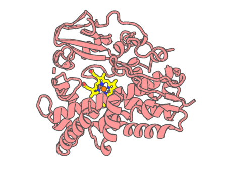 |
Experimental method | X-RAY DIFFRACTION |
|---|---|---|
| Structural data | unavailable | |
| Non-standard amino acids | None | |
| Missing residues/ Structural gap | Exists / Exists | |
| Resolution | 2.36 Å | |
| Deposition date | 2012-03-09 |