
Number of data: 7660
Dataset: all data
| pdb_00004kew | structure of the A82F BM3 heme domain in complex with omeprazole | ||
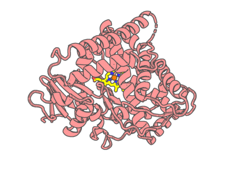 |
Experimental method | X-RAY DIFFRACTION |
|---|---|---|
| Structural data | available | |
| Non-standard amino acids | None | |
| Missing residues/ Structural gap | Exists / Exists | |
| Resolution | 1.89 Å | |
| Deposition date | 2013-04-26 |
| pdb_00004key | Structure of P450 BM3 A82F F87V in complex with omeprazole | ||
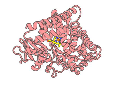 |
Experimental method | X-RAY DIFFRACTION |
|---|---|---|
| Structural data | available | |
| Non-standard amino acids | None | |
| Missing residues/ Structural gap | Exists / Exists | |
| Resolution | 2.05 Å | |
| Deposition date | 2013-04-26 |
| pdb_00004kf0 | Structure of the A82F P450 BM3 heme domain | ||
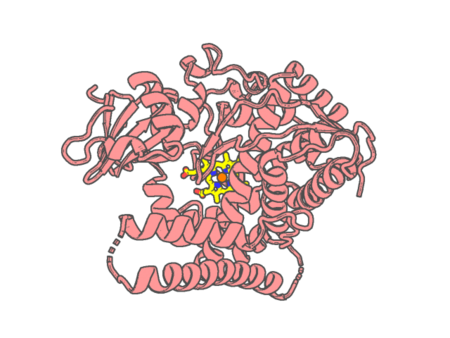 |
Experimental method | X-RAY DIFFRACTION |
|---|---|---|
| Structural data | available | |
| Non-standard amino acids | None | |
| Missing residues/ Structural gap | Exists / Exists | |
| Resolution | 1.45 Å | |
| Deposition date | 2013-04-26 |
| pdb_00004kf2 | Structure of the P4509 BM3 A82F F87V heme domain | ||
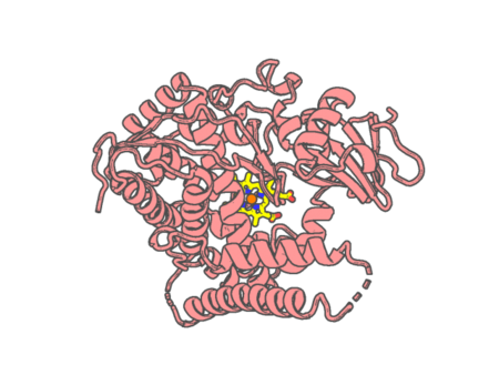 |
Experimental method | X-RAY DIFFRACTION |
|---|---|---|
| Structural data | available | |
| Non-standard amino acids | None | |
| Missing residues/ Structural gap | Exists / Exists | |
| Resolution | 1.82 Å | |
| Deposition date | 2013-04-26 |
| pdb_00004kjt | Structure of the L100F MUTANT OF DEHALOPEROXIDASE-HEMOGLOBIN A FROM AMPHITRITE ORNATA WITH OXYGEN | ||
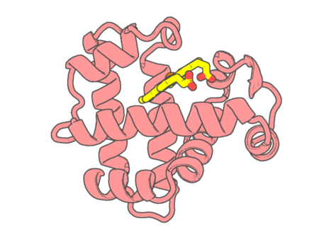 |
Experimental method | X-RAY DIFFRACTION |
|---|---|---|
| Structural data | available | |
| Non-standard amino acids | None | |
| Missing residues/ Structural gap | None / None | |
| Resolution | 1.44 Å | |
| Deposition date | 2013-05-03 |
| pdb_00004kky | Crystal structure of N-(1-Pyrene)acetamide labeled P450cam in substrate bound form. | ||
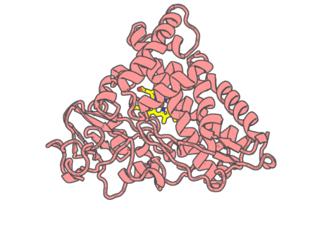 |
Experimental method | X-RAY DIFFRACTION |
|---|---|---|
| Structural data | available | |
| Non-standard amino acids | None | |
| Missing residues/ Structural gap | None / None | |
| Resolution | 2 Å | |
| Deposition date | 2013-05-06 |
| pdb_00004klc | E343D/F110A Double mutant of human ferrochelatase | ||
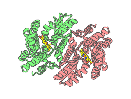 |
Experimental method | X-RAY DIFFRACTION |
|---|---|---|
| Structural data | available | |
| Non-standard amino acids | None | |
| Missing residues/ Structural gap | None / None | |
| Resolution | 2.4 Å | |
| Deposition date | 2013-05-07 |
| pdb_00004klr | E343Q variant of human ferrochelatase | ||
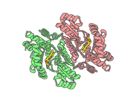 |
Experimental method | X-RAY DIFFRACTION |
|---|---|---|
| Structural data | available | |
| Non-standard amino acids | None | |
| Missing residues/ Structural gap | None / None | |
| Resolution | 2.18 Å | |
| Deposition date | 2013-05-07 |
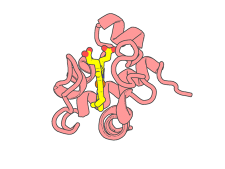
| pdb_00004kmg | Crystal structure of cytochrome c6B from Synechococcus sp. WH8102 | ||
 |
Experimental method | X-RAY DIFFRACTION |
|---|---|---|
| Structural data | available | |
| Non-standard amino acids | None | |
| Missing residues/ Structural gap | None / None | |
| Resolution | 1.4 Å | |
| Deposition date | 2013-05-08 |
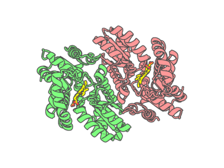
| pdb_00004kmm | M76H variant of human ferrochelatase | ||
 |
Experimental method | X-RAY DIFFRACTION |
|---|---|---|
| Structural data | available | |
| Non-standard amino acids | None | |
| Missing residues/ Structural gap | None / None | |
| Resolution | 2.6 Å | |
| Deposition date | 2013-05-08 |
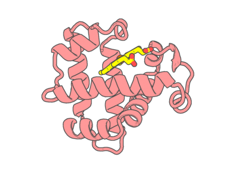
| pdb_00004kmv | Structure of the L100F MUTANT OF DEHALOPEROXIDASE-HEMOGLOBIN A FROM AMPHITRITE ORNATA WITH 2,4,6-TRICHLOROPHENOL | ||
 |
Experimental method | X-RAY DIFFRACTION |
|---|---|---|
| Structural data | available | |
| Non-standard amino acids | None | |
| Missing residues/ Structural gap | None / None | |
| Resolution | 1.44 Å | |
| Deposition date | 2013-05-08 |
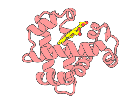
| pdb_00004kmw | Structure of the Y34N MUTANT OF DEHALOPEROXIDASE-HEMOGLOBIN A FROM AMPHITRITE ORNATA WITH 2,4,6-TRICHLOROPHENOL | ||
 |
Experimental method | X-RAY DIFFRACTION |
|---|---|---|
| Structural data | available | |
| Non-standard amino acids | None | |
| Missing residues/ Structural gap | None / None | |
| Resolution | 1.79 Å | |
| Deposition date | 2013-05-08 |
| pdb_00004kn3 | Structure of the Y34NS91G double mutant of Dehaloperoxidase from Amphitrite ornata with 2,4,6-trichlorophenol | ||
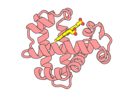 |
Experimental method | X-RAY DIFFRACTION |
|---|---|---|
| Structural data | available | |
| Non-standard amino acids | None | |
| Missing residues/ Structural gap | None / None | |
| Resolution | 1.78 Å | |
| Deposition date | 2013-05-08 |
| pdb_00004kpa | Crystal structure of cytochrome P450 BM-3 in complex with N-palmitoylglycine (NPG) | ||
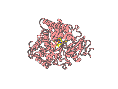 |
Experimental method | X-RAY DIFFRACTION |
|---|---|---|
| Structural data | available | |
| Non-standard amino acids | None | |
| Missing residues/ Structural gap | Exists / Exists | |
| Resolution | 2 Å | |
| Deposition date | 2013-05-13 |
| pdb_00004kpb | Crystal structure of cytochrome P450 BM-3 R47E mutant | ||
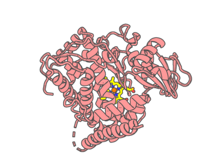 |
Experimental method | X-RAY DIFFRACTION |
|---|---|---|
| Structural data | available | |
| Non-standard amino acids | None | |
| Missing residues/ Structural gap | Exists / Exists | |
| Resolution | 2.1 Å | |
| Deposition date | 2013-05-13 |
| pdb_00004kq8 | Structure of Recombinant Human Cytochrome P450 Aromatase | ||
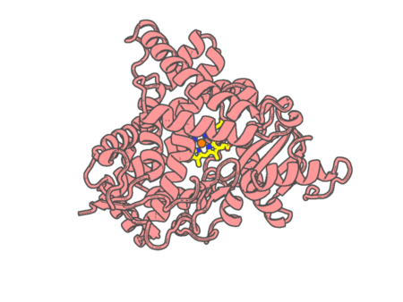 |
Experimental method | X-RAY DIFFRACTION |
|---|---|---|
| Structural data | available | |
| Non-standard amino acids | None | |
| Missing residues/ Structural gap | None / None | |
| Resolution | 3.29 Å | |
| Deposition date | 2013-05-14 |
| pdb_00004ksz | Crystal structure of bovine lactoperoxidase complexed with cystiene at 1.98A resolution | ||
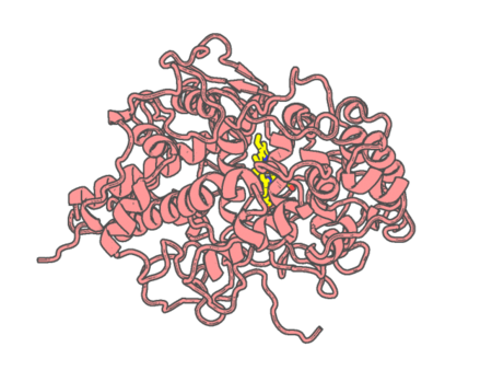 |
Experimental method | X-RAY DIFFRACTION |
|---|---|---|
| Structural data | available | |
| Non-standard amino acids | Included | |
| Missing residues/ Structural gap | None / None | |
| Resolution | 1.98 Å | |
| Deposition date | 2013-05-19 |
| pdb_00004ktf | Crystal structure of Mycobacterium tuberculosis CYP121 in complex with 4,4'-(3-amino-1H-pyrazole-4,5-diyl)diphenol | ||
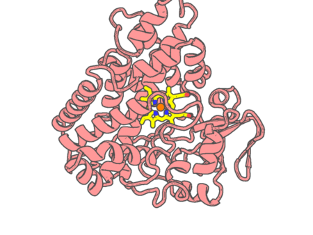 |
Experimental method | X-RAY DIFFRACTION |
|---|---|---|
| Structural data | available | |
| Non-standard amino acids | None | |
| Missing residues/ Structural gap | Exists / None | |
| Resolution | 1.35 Å | |
| Deposition date | 2013-05-20 |
| pdb_00004ktj | Crystal structure of Mycobacterium tuberculosis CYP121 in complex with 4-(3-amino-1H-pyrazol-4-yl)phenol | ||
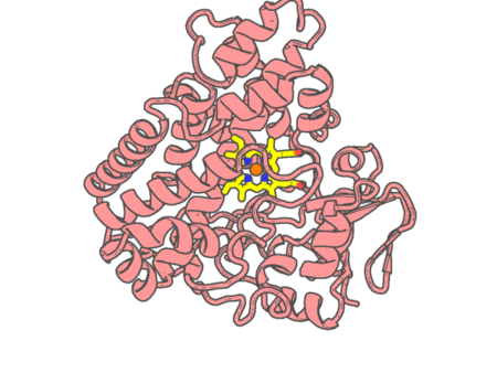 |
Experimental method | X-RAY DIFFRACTION |
|---|---|---|
| Structural data | available | |
| Non-standard amino acids | None | |
| Missing residues/ Structural gap | None / None | |
| Resolution | 1.35 Å | |
| Deposition date | 2013-05-20 |
| pdb_00004ktk | Crystal structure of Mycobacterium tuberculosis CYP121 in complex with 4-(3-amino-4-(4-hydroxyphenyl)-1H-pyrazol-5-yl)benzene-1,3-diol | ||
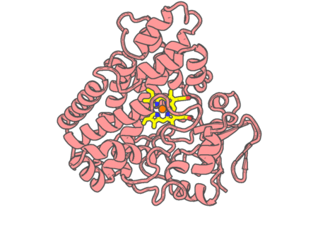 |
Experimental method | X-RAY DIFFRACTION |
|---|---|---|
| Structural data | available | |
| Non-standard amino acids | None | |
| Missing residues/ Structural gap | Exists / None | |
| Resolution | 1.4 Å | |
| Deposition date | 2013-05-20 |