Number of data: 7660
Dataset: all data
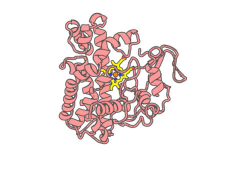 |
Experimental method |
X-RAY DIFFRACTION |
| Structural data |
available |
| Non-standard amino acids |
None |
| Missing residues/ Structural gap |
Exists / Exists |
| Resolution |
1.9 Å |
| Deposition date |
2014-06-26 |
| pdb_00004tx3 |
Complex of the X-domain and OxyB from Teicoplanin Biosynthesis
|
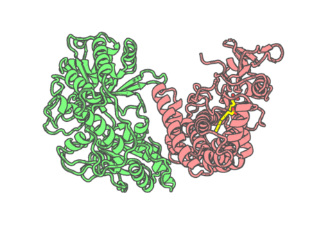 |
Experimental method |
X-RAY DIFFRACTION |
| Structural data |
available |
| Non-standard amino acids |
None |
| Missing residues/ Structural gap |
Exists / Exists |
| Resolution |
2.5 Å |
| Deposition date |
2014-07-02 |
| pdb_00004tyx |
Structure of aquoferric sperm whale myoglobin L29H/F33Y/F43H/S92A mutant
|
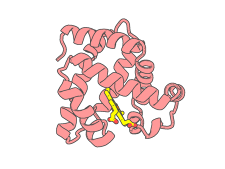 |
Experimental method |
X-RAY DIFFRACTION |
| Structural data |
available |
| Non-standard amino acids |
None |
| Missing residues/ Structural gap |
None / None |
| Resolution |
1.64 Å |
| Deposition date |
2014-07-09 |
| pdb_00004u3f |
Cytochrome bc1 complex from chicken with designed inhibitor bound
|
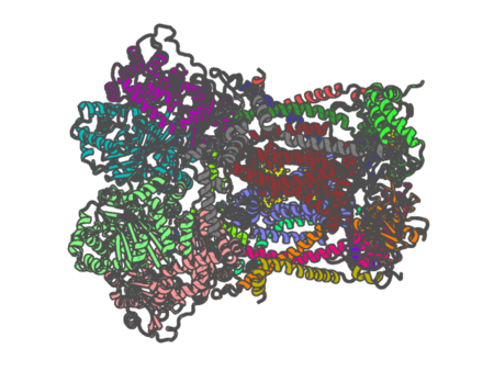 |
Experimental method |
X-RAY DIFFRACTION |
| Structural data |
available |
| Non-standard amino acids |
Included |
| Missing residues/ Structural gap |
Exists / None |
| Resolution |
3.2312 Å |
| Deposition date |
2014-07-21 |
| pdb_00004u72 |
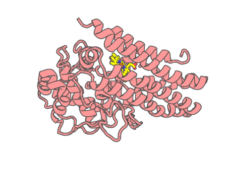
Crystal structure of 4-phenylimidazole bound form of human indoleamine 2,3-dioxygenase (A260G mutant)
|
 |
Experimental method |
X-RAY DIFFRACTION |
| Structural data |
available |
| Non-standard amino acids |
None |
| Missing residues/ Structural gap |
Exists / Exists |
| Resolution |
2 Å |
| Deposition date |
2014-07-30 |
| pdb_00004u74 |
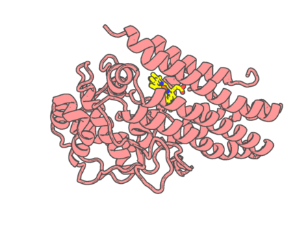
Crystal structure of 4-phenylimidazole bound form of human indoleamine 2,3-dioxygenase (G262A mutant)
|
 |
Experimental method |
X-RAY DIFFRACTION |
| Structural data |
available |
| Non-standard amino acids |
None |
| Missing residues/ Structural gap |
Exists / Exists |
| Resolution |
2.31 Å |
| Deposition date |
2014-07-30 |
| pdb_00004u8u |
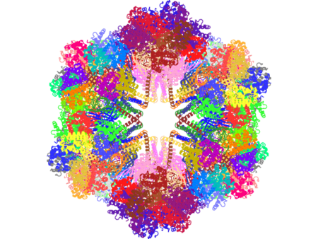
The Crystallographic structure of the giant hemoglobin from Glossoscolex paulistus at 3.2 A resolution.
|
 |
Experimental method |
X-RAY DIFFRACTION |
| Structural data |
available |
| Non-standard amino acids |
None |
| Missing residues/ Structural gap |
None / None |
| Resolution |
3.2 Å |
| Deposition date |
2014-08-04 |
| pdb_00004u99 |
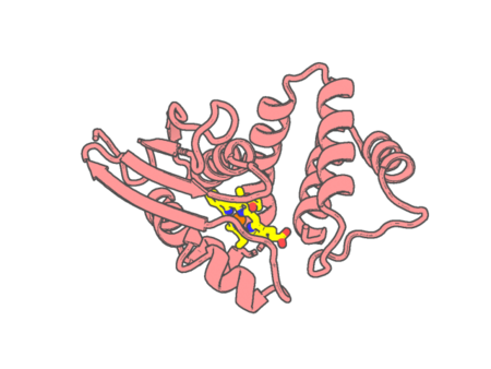
Crystal structure of an H-NOX protein from S. oneidensis in the Fe(II) ligation state, Q154A/Q155A/K156A mutant
|
 |
Experimental method |
X-RAY DIFFRACTION |
| Structural data |
available |
| Non-standard amino acids |
None |
| Missing residues/ Structural gap |
None / None |
| Resolution |
2 Å |
| Deposition date |
2014-08-05 |
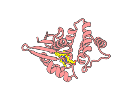
| pdb_00004u9b |
Crystal structure of an H-NOX protein from S. oneidensis in the Fe(II)NO ligation state
|
 |
Experimental method |
X-RAY DIFFRACTION |
| Structural data |
available |
| Non-standard amino acids |
None |
| Missing residues/ Structural gap |
None / None |
| Resolution |
1.65 Å |
| Deposition date |
2014-08-05 |
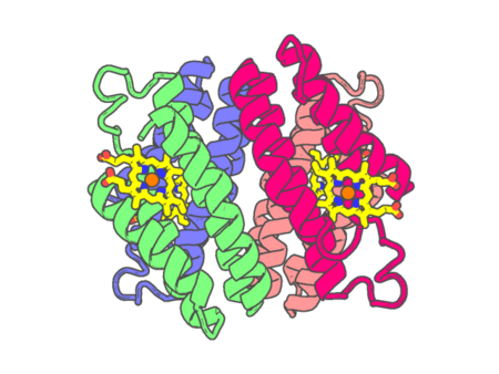
| pdb_00004u9d |
Crystal Structure of the Zn-directed tetramer of the engineered cyt cb562 variant, AB3
|
 |
Experimental method |
X-RAY DIFFRACTION |
| Structural data |
available |
| Non-standard amino acids |
None |
| Missing residues/ Structural gap |
None / None |
| Resolution |
2.5 Å |
| Deposition date |
2014-08-05 |
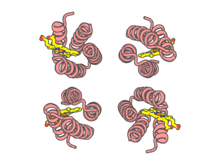
| pdb_00004u9e |
Crystal structure of the Zn-directed tetramer of the engineered cyt cb562 variant, A104/57G AB3
|
 |
Experimental method |
X-RAY DIFFRACTION |
| Structural data |
available |
| Non-standard amino acids |
None |
| Missing residues/ Structural gap |
Exists / Exists |
| Resolution |
2.8 Å |
| Deposition date |
2014-08-06 |
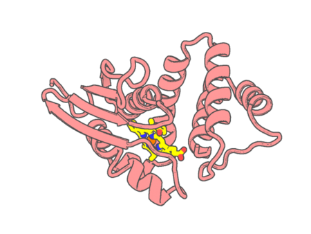
| pdb_00004u9g |
Crystal structure of an H-NOX protein from S. oneidensis in the Fe(II)CO ligation state, Q154A/Q155A/K156A mutant
|
 |
Experimental method |
X-RAY DIFFRACTION |
| Structural data |
available |
| Non-standard amino acids |
None |
| Missing residues/ Structural gap |
None / None |
| Resolution |
2.25 Å |
| Deposition date |
2014-08-06 |
| pdb_00004uax |
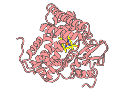
X-ray crystal structure of ligand free CYP142A2 from Mycobacterium smegmatis
|
 |
Experimental method |
X-RAY DIFFRACTION |
| Structural data |
available |
| Non-standard amino acids |
None |
| Missing residues/ Structural gap |
None / None |
| Resolution |
1.78 Å |
| Deposition date |
2014-08-11 |
| pdb_00004ub6 |
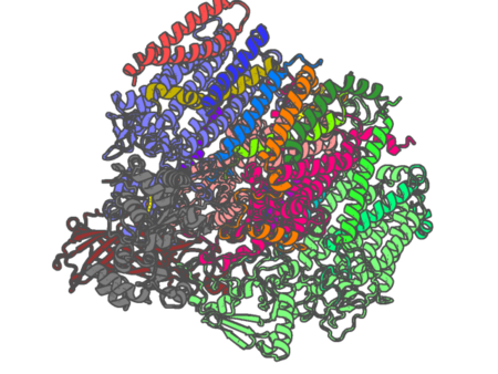
Native structure of photosystem II (dataset-1) by a femtosecond X-ray laser
|
 |
Experimental method |
X-RAY DIFFRACTION |
| Structural data |
available |
| Non-standard amino acids |
Included |
| Missing residues/ Structural gap |
None / None |
| Resolution |
1.95 Å |
| Deposition date |
2014-08-12 |
| pdb_00004ub8 |
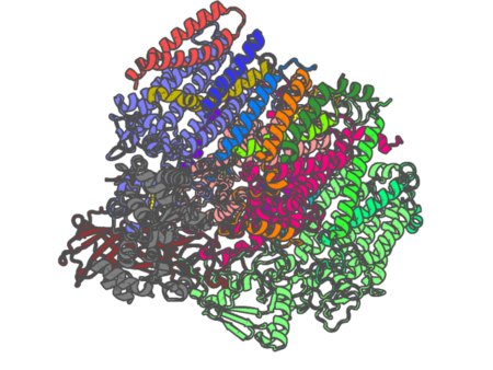
Native structure of photosystem II (dataset-2) by a femtosecond X-ray laser
|
 |
Experimental method |
X-RAY DIFFRACTION |
| Structural data |
available |
| Non-standard amino acids |
Included |
| Missing residues/ Structural gap |
None / None |
| Resolution |
1.95 Å |
| Deposition date |
2014-08-12 |
| pdb_00004ubs |
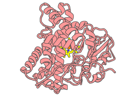
The crystal structure of cytochrome P450 105D7 from Streptomyces avermitilis in complex with Diclofenac
|
 |
Experimental method |
X-RAY DIFFRACTION |
| Structural data |
available |
| Non-standard amino acids |
None |
| Missing residues/ Structural gap |
None / None |
| Resolution |
2.2 Å |
| Deposition date |
2014-08-13 |
| pdb_00004uch |
Structure of human nNOS R354A G357D mutant heme domain in complex with 3-(((2-((2-(1H-IMIDAZOL-1-YL)PYRIMIDIN-4-YL)ETHYL)AMINO)METHYL) BENZONITRILE
|
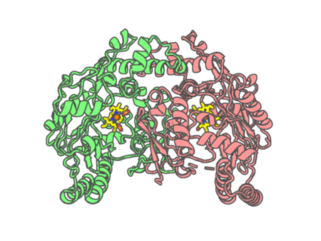 |
Experimental method |
X-RAY DIFFRACTION |
| Structural data |
available |
| Non-standard amino acids |
None |
| Missing residues/ Structural gap |
Exists / Exists |
| Resolution |
2.2 Å |
| Deposition date |
2014-12-03 |
| pdb_00004ug5 |
Structure of Bacillus subtilis Nitric Oxide Synthase in complex with 6-(2-(5-(2-(2-amino-6-methylpyridin-4-yl)ethyl)pyridin-3-yl)ethyl)-4- methylpyridin-2-amine
|
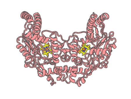 |
Experimental method |
X-RAY DIFFRACTION |
| Structural data |
available |
| Non-standard amino acids |
None |
| Missing residues/ Structural gap |
None / None |
| Resolution |
2.352 Å |
| Deposition date |
2015-03-22 |
| pdb_00004ug6 |
Structure of Bacillus subtilis Nitric Oxide Synthase in complex with 6,6'-(pyridine-3,5-diyldiethane-2,1-diyl)bis(4-methylpyridin-2-amine)
|
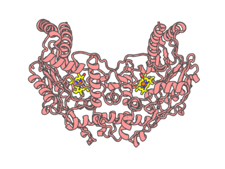 |
Experimental method |
X-RAY DIFFRACTION |
| Structural data |
available |
| Non-standard amino acids |
None |
| Missing residues/ Structural gap |
None / None |
| Resolution |
1.81 Å |
| Deposition date |
2015-03-22 |
| pdb_00004ug7 |
Structure of Bacillus subtilis Nitric Oxide Synthase in complex with 3,5-bis(2-(6-amino-4-methylpyridin-2-yl)ethyl)benzonitrile
|
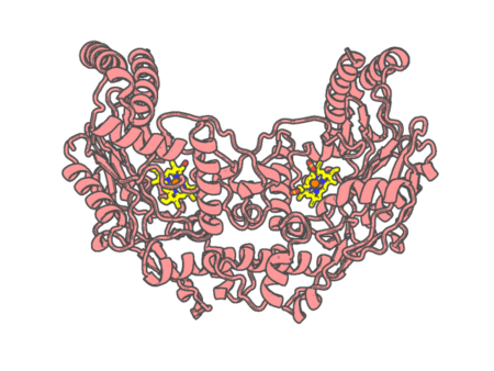 |
Experimental method |
X-RAY DIFFRACTION |
| Structural data |
available |
| Non-standard amino acids |
None |
| Missing residues/ Structural gap |
None / None |
| Resolution |
1.76 Å |
| Deposition date |
2015-03-22 |
