
Number of data: 7660
Dataset: all data
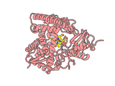
| pdb_00006mi0 | Crystal structure of the P450 domain of the CYP51-ferredoxin fusion protein from Methylococcus capsulatus, ligand-free state | ||
 |
Experimental method | X-RAY DIFFRACTION |
|---|---|---|
| Structural data | available | |
| Non-standard amino acids | None | |
| Missing residues/ Structural gap | None / None | |
| Resolution | 2.73 Å | |
| Deposition date | 2018-09-18 |
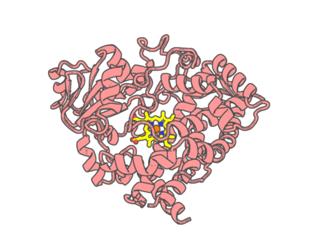
| pdb_00006mjm | Substrate Free Cytochrome P450 3A5 (CYP3A5) | ||
 |
Experimental method | X-RAY DIFFRACTION |
|---|---|---|
| Structural data | available | |
| Non-standard amino acids | None | |
| Missing residues/ Structural gap | Exists / Exists | |
| Resolution | 2.2 Å | |
| Deposition date | 2018-09-21 |
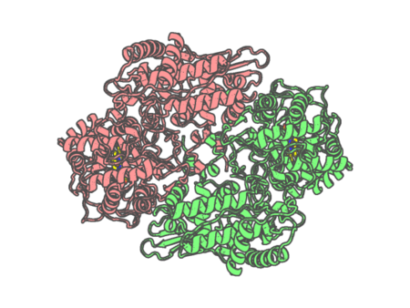
| pdb_00006mpy | B. pseudomallei KatG crystallized in the presence of benzoyl hydrazide | ||
 |
Experimental method | X-RAY DIFFRACTION |
|---|---|---|
| Structural data | available | |
| Non-standard amino acids | None | |
| Missing residues/ Structural gap | None / None | |
| Resolution | 2 Å | |
| Deposition date | 2018-10-09 |
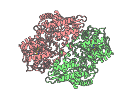
| pdb_00006mq0 | B. pseudomallei KatG crystallized in the presence of furoyl hydrazide | ||
 |
Experimental method | X-RAY DIFFRACTION |
|---|---|---|
| Structural data | available | |
| Non-standard amino acids | None | |
| Missing residues/ Structural gap | None / None | |
| Resolution | 1.9 Å | |
| Deposition date | 2018-10-09 |
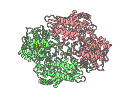
| pdb_00006mq1 | B. pseudomallei KatG crystalized in the presence of ABTS | ||
 |
Experimental method | X-RAY DIFFRACTION |
|---|---|---|
| Structural data | available | |
| Non-standard amino acids | None | |
| Missing residues/ Structural gap | None / None | |
| Resolution | 2.5 Å | |
| Deposition date | 2018-10-09 |
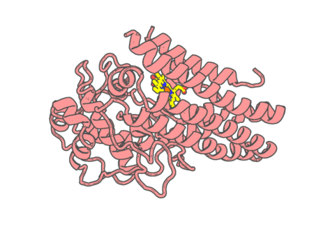
| pdb_00006mq6 | Mapping the binding trajectory of a suicide inhibitor in human indoleamine 2,3-dioxygenase 1 | ||
 |
Experimental method | X-RAY DIFFRACTION |
|---|---|---|
| Structural data | available | |
| Non-standard amino acids | None | |
| Missing residues/ Structural gap | Exists / Exists | |
| Resolution | 3.05 Å | |
| Deposition date | 2018-10-09 |
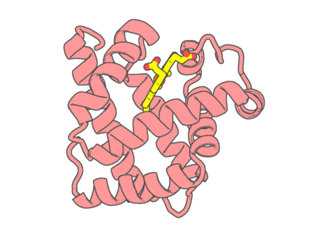
| pdb_00006mv0 | CO-bound Sperm Whale Myoglobin, room temperature structure solved by serial 5degree oscillation crystallography | ||
 |
Experimental method | X-RAY DIFFRACTION |
|---|---|---|
| Structural data | available | |
| Non-standard amino acids | None | |
| Missing residues/ Structural gap | None / None | |
| Resolution | 1.97 Å | |
| Deposition date | 2018-10-24 |
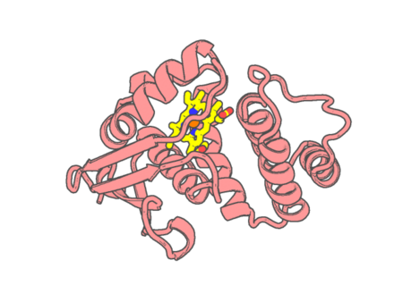
| pdb_00006mx5 | Crystal structure of H-NOX protein from Nostoc sp. | ||
 |
Experimental method | X-RAY DIFFRACTION |
|---|---|---|
| Structural data | available | |
| Non-standard amino acids | None | |
| Missing residues/ Structural gap | None / None | |
| Resolution | 2.35 Å | |
| Deposition date | 2018-10-30 |
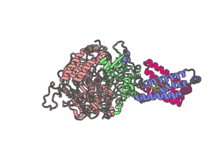
| pdb_00006myo | Avian mitochondrial complex II with flutolanyl bound | ||
 |
Experimental method | X-RAY DIFFRACTION |
|---|---|---|
| Structural data | available | |
| Non-standard amino acids | Included | |
| Missing residues/ Structural gap | None / None | |
| Resolution | 2.2 Å | |
| Deposition date | 2018-11-02 |
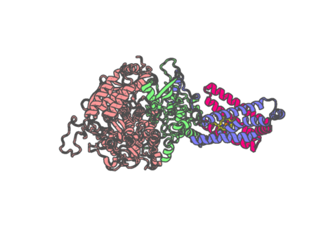
| pdb_00006myp | Avian mitochondrial complex II with TTFA (thenoyltrifluoroacetone) bound | ||
 |
Experimental method | X-RAY DIFFRACTION |
|---|---|---|
| Structural data | available | |
| Non-standard amino acids | Included | |
| Missing residues/ Structural gap | None / None | |
| Resolution | 2.1 Å | |
| Deposition date | 2018-11-02 |
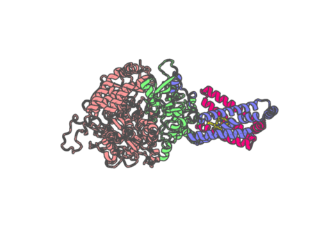
| pdb_00006myq | Avian mitochondrial complex II with ferulenol bound | ||
 |
Experimental method | X-RAY DIFFRACTION |
|---|---|---|
| Structural data | available | |
| Non-standard amino acids | Included | |
| Missing residues/ Structural gap | None / None | |
| Resolution | 1.97 Å | |
| Deposition date | 2018-11-02 |
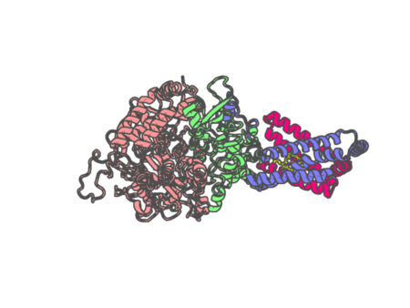
| pdb_00006myr | Avian mitochondrial complex II with thiapronil bound | ||
 |
Experimental method | X-RAY DIFFRACTION |
|---|---|---|
| Structural data | available | |
| Non-standard amino acids | None | |
| Missing residues/ Structural gap | None / None | |
| Resolution | 2.15 Å | |
| Deposition date | 2018-11-02 |
| pdb_00006mys | Avian mitochondrial complex II with Atpenin A5 bound, sidechain outside | ||
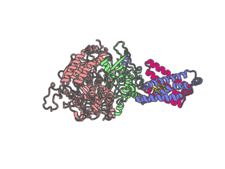 |
Experimental method | X-RAY DIFFRACTION |
|---|---|---|
| Structural data | available | |
| Non-standard amino acids | Included | |
| Missing residues/ Structural gap | None / None | |
| Resolution | 2.37 Å | |
| Deposition date | 2018-11-02 |
| pdb_00006myt | Avian mitochondrial complex II with Atpenin A5 bound, sidechain in pocket | ||
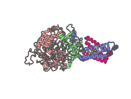 |
Experimental method | X-RAY DIFFRACTION |
|---|---|---|
| Structural data | available | |
| Non-standard amino acids | Included | |
| Missing residues/ Structural gap | None / None | |
| Resolution | 2.27 Å | |
| Deposition date | 2018-11-02 |
| pdb_00006myu | Avian mitochondrial complex II crystallized in the presence of HQNO | ||
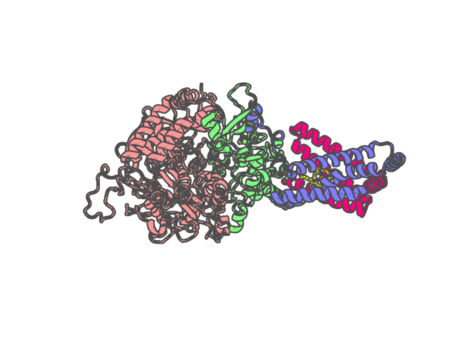 |
Experimental method | X-RAY DIFFRACTION |
|---|---|---|
| Structural data | available | |
| Non-standard amino acids | Included | |
| Missing residues/ Structural gap | None / None | |
| Resolution | 1.97 Å | |
| Deposition date | 2018-11-02 |
| pdb_00006n02 | CO-bound Sperm Whale Myoglobin, room temperature structure, first 2 degrees of 5 degree total oscillation | ||
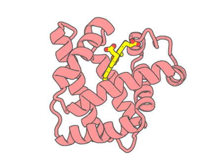 |
Experimental method | X-RAY DIFFRACTION |
|---|---|---|
| Structural data | available | |
| Non-standard amino acids | None | |
| Missing residues/ Structural gap | None / None | |
| Resolution | 2 Å | |
| Deposition date | 2018-11-06 |
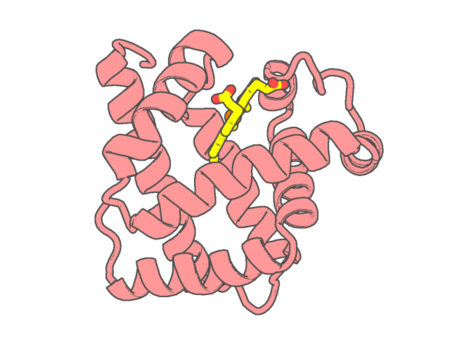
| pdb_00006n03 | CO-bound Sperm Whale Myoglobin, room temperature structure, last 2 degrees of 5 degree total oscillation and 160 kGy dose | ||
 |
Experimental method | X-RAY DIFFRACTION |
|---|---|---|
| Structural data | available | |
| Non-standard amino acids | None | |
| Missing residues/ Structural gap | None / None | |
| Resolution | 2.1 Å | |
| Deposition date | 2018-11-06 |
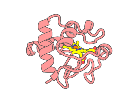
| pdb_00006n1o | Oxidized rat cytochrome c mutant (S47E) | ||
 |
Experimental method | X-RAY DIFFRACTION |
|---|---|---|
| Structural data | available | |
| Non-standard amino acids | None | |
| Missing residues/ Structural gap | None / None | |
| Resolution | 1.55 Å | |
| Deposition date | 2018-11-09 |
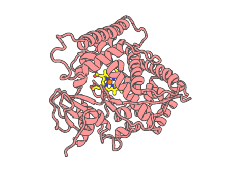
| pdb_00006n6q | Crystal structure of a Cytochrome P450 (CYP102L1) | ||
 |
Experimental method | X-RAY DIFFRACTION |
|---|---|---|
| Structural data | available | |
| Non-standard amino acids | None | |
| Missing residues/ Structural gap | None / None | |
| Resolution | 2.5 Å | |
| Deposition date | 2018-11-26 |
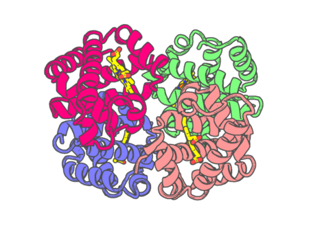
| pdb_00006nbc | human methemoglobin state 1 determined using single-particle cryo-EM at 200 keV | ||
 |
Experimental method | ELECTRON MICROSCOPY |
|---|---|---|
| Structural data | available | |
| Non-standard amino acids | None | |
| Missing residues/ Structural gap | None / None | |
| Resolution | None | |
| Deposition date | 2018-12-06 |