
Number of data: 3514
Dataset: (without mutations)
| pdb_000021rt | Cryo-EM structure of mouse myeloperoxidase in complex with Fab fragments of antibodies mAb-A46 and mAb-B88 | ||
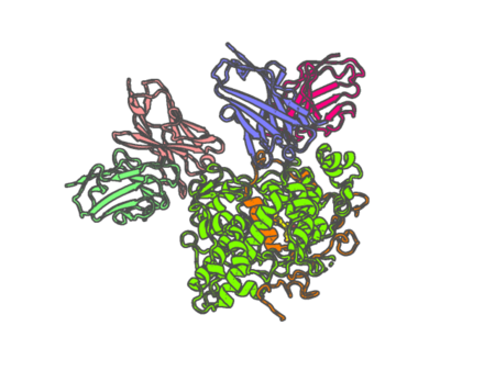 |
Experimental method | ELECTRON MICROSCOPY |
|---|---|---|
| Structural data | available | |
| Non-standard amino acids | None | |
| Missing residues/ Structural gap | Exists / Exists | |
| Resolution | None | |
| Deposition date | 2025-12-23 |
| pdb_000021wv | In situ structure of the C2S2M2L4-type PSII-LHCII supercomplex from Oryza sativa | ||
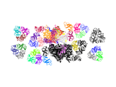 |
Experimental method | ELECTRON MICROSCOPY |
|---|---|---|
| Structural data | available | |
| Non-standard amino acids | None | |
| Missing residues/ Structural gap | Exists / Exists | |
| Resolution | 2.89 Å | |
| Deposition date | 2026-01-01 |
| pdb_000022yx | RufO homolog binding with Nle-Arg-Tyr-Leu-His from Nonomuraea solani | ||
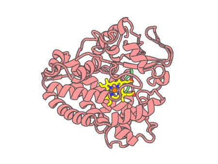 |
Experimental method | X-RAY DIFFRACTION |
|---|---|---|
| Structural data | available | |
| Non-standard amino acids | None | |
| Missing residues/ Structural gap | Exists / Exists | |
| Resolution | 1.59 Å | |
| Deposition date | 2026-01-28 |
| pdb_000023ub | Structure of the CYP102A1 Heme Domain with 2-(undecylcarbamoyl)benzoic acid | ||
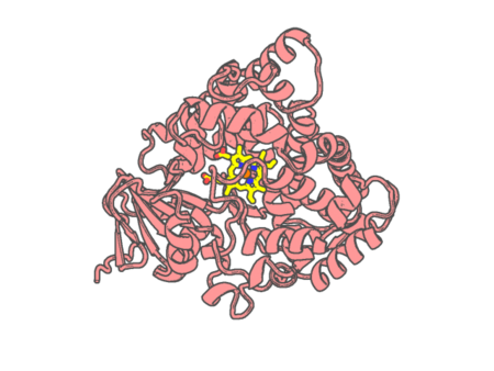 |
Experimental method | X-RAY DIFFRACTION |
|---|---|---|
| Structural data | available | |
| Non-standard amino acids | None | |
| Missing residues/ Structural gap | None / None | |
| Resolution | 1.63 Å | |
| Deposition date | 2026-02-17 |
| pdb_000023uh | Structure of the CYP102A1 Heme Domain with 2-(undecylcarbamoyl)benzoic acid and benzene | ||
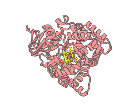 |
Experimental method | X-RAY DIFFRACTION |
|---|---|---|
| Structural data | available | |
| Non-standard amino acids | None | |
| Missing residues/ Structural gap | None / None | |
| Resolution | 1.48 Å | |
| Deposition date | 2026-02-18 |
| pdb_0000256b | IMPROVEMENT OF THE 2.5 ANGSTROMS RESOLUTION MODEL OF CYTOCHROME B562 BY REDETERMINING THE PRIMARY STRUCTURE AND USING MOLECULAR GRAPHICS | ||
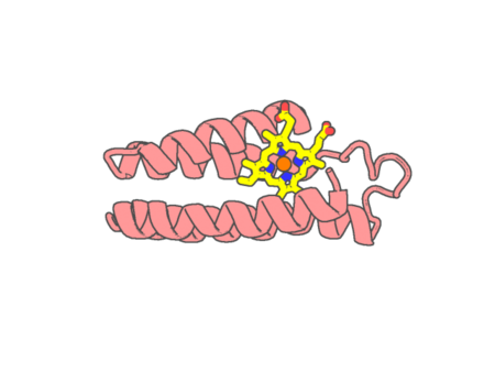 |
Experimental method | X-RAY DIFFRACTION |
|---|---|---|
| Structural data | available | |
| Non-standard amino acids | None | |
| Missing residues/ Structural gap | None / None | |
| Resolution | 1.4 Å | |
| Deposition date | 1990-01-16 |
| pdb_000028od | Crystal structure of C35 bound to Hem | ||
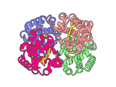 |
Experimental method | X-RAY DIFFRACTION |
|---|---|---|
| Structural data | available | |
| Non-standard amino acids | None | |
| Missing residues/ Structural gap | None / None | |
| Resolution | 1.73 Å | |
| Deposition date | 2026-02-10 |
| pdb_000028pb | Crystal structure of CbcA periplasmic domain from Geobacter sulfurreducens | ||
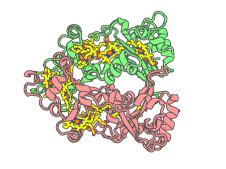 |
Experimental method | X-RAY DIFFRACTION |
|---|---|---|
| Structural data | available | |
| Non-standard amino acids | None | |
| Missing residues/ Structural gap | None / None | |
| Resolution | 1.91 Å | |
| Deposition date | 2026-02-12 |
| pdb_000028pn | CryoEM structure of native quinol dependent Nitric Oxide Reductase at pH 8.0 on gold grid. | ||
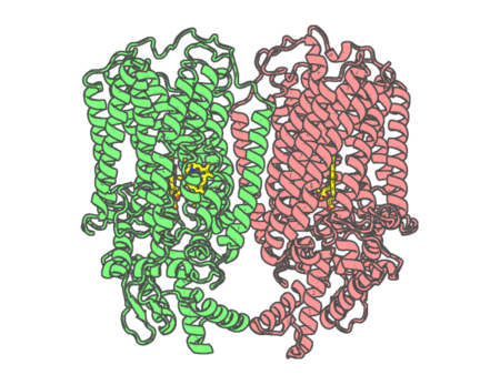 |
Experimental method | ELECTRON MICROSCOPY |
|---|---|---|
| Structural data | unavailable | |
| Non-standard amino acids | None | |
| Missing residues/ Structural gap | Exists / Exists | |
| Resolution | 2.7 Å | |
| Deposition date | 2026-02-12 |
| pdb_000028wu | Crystal Structure of Catalase HPII (KatE) from Escherichia coli | ||
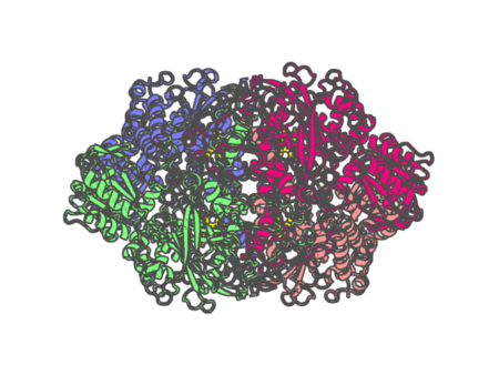 |
Experimental method | X-RAY DIFFRACTION |
|---|---|---|
| Structural data | available | |
| Non-standard amino acids | None | |
| Missing residues/ Structural gap | None / None | |
| Resolution | 2.5 Å | |
| Deposition date | 2026-02-25 |
| pdb_000029to | Structure of monomerized and de-glycosylated native leukocyte myeloperoxidase in complex with the Staphylococcal Peroxidase Inhibitor SPIN | ||
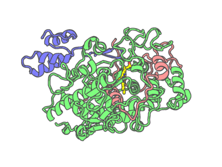 |
Experimental method | X-RAY DIFFRACTION |
|---|---|---|
| Structural data | available | |
| Non-standard amino acids | Included | |
| Missing residues/ Structural gap | Exists / Exists | |
| Resolution | 1.44 Å | |
| Deposition date | 2026-04-08 |
| pdb_000029wj | Structure of monomerized native leukocyte myeloperoxidase in complex with the Staphylococcal Peroxidase Inhibitor SPIN | ||
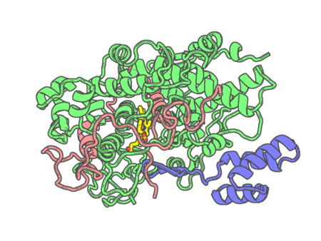 |
Experimental method | X-RAY DIFFRACTION |
|---|---|---|
| Structural data | available | |
| Non-standard amino acids | Included | |
| Missing residues/ Structural gap | None / None | |
| Resolution | 2.55 Å | |
| Deposition date | 2026-04-10 |
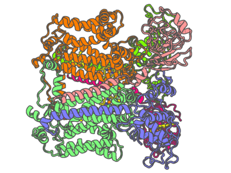
| pdb_000029xz | Cryo-EM structure of Rhodobacter capsulatus cytochrome bc1 dimer at 2.16 A resolution with water molecules and quinone in the quinone reduction site | ||
 |
Experimental method | ELECTRON MICROSCOPY |
|---|---|---|
| Structural data | available | |
| Non-standard amino acids | None | |
| Missing residues/ Structural gap | Exists / Exists | |
| Resolution | None | |
| Deposition date | 2026-04-14 |
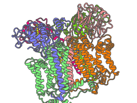
| pdb_000029ya | Cryo-EM structure of Rhodobacter capsulatus cytochrome bc1 dimer with one Rieske protein in the c position and one in the b position and quinone in the quinone oxidation site | ||
 |
Experimental method | ELECTRON MICROSCOPY |
|---|---|---|
| Structural data | available | |
| Non-standard amino acids | None | |
| Missing residues/ Structural gap | Exists / Exists | |
| Resolution | 2.69 Å | |
| Deposition date | 2026-04-14 |
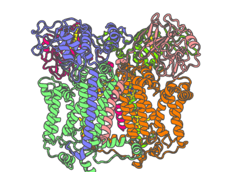
| pdb_000029yb | Cryo-EM structure of Rhodobacter capsulatus cytochrome bc1 dimer with both Rieske proteins in the b position | ||
 |
Experimental method | ELECTRON MICROSCOPY |
|---|---|---|
| Structural data | available | |
| Non-standard amino acids | None | |
| Missing residues/ Structural gap | Exists / Exists | |
| Resolution | None | |
| Deposition date | 2026-04-14 |
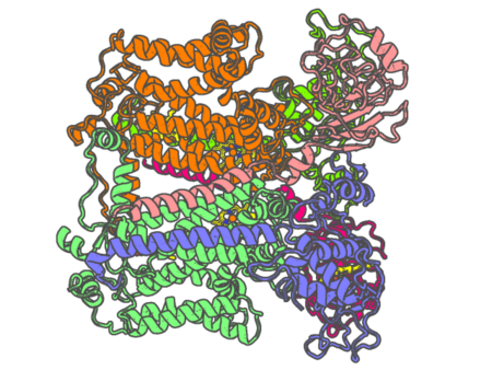
| pdb_000029yc | Cryo-EM structure of Rhodobacter capsulatus cytochrome bc1 dimer with both Rieske proteins in the c position | ||
 |
Experimental method | ELECTRON MICROSCOPY |
|---|---|---|
| Structural data | available | |
| Non-standard amino acids | None | |
| Missing residues/ Structural gap | Exists / Exists | |
| Resolution | 2.43 Å | |
| Deposition date | 2026-04-14 |
| pdb_000029yd | Cryo-EM structure of Rhodobacter capsulatus cytochrome bc1 dimer with one Rieske protein in the c position and one in an intermediate position | ||
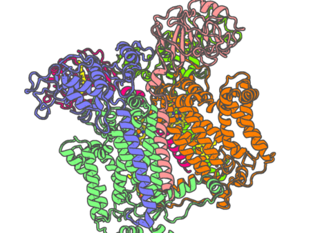 |
Experimental method | ELECTRON MICROSCOPY |
|---|---|---|
| Structural data | available | |
| Non-standard amino acids | None | |
| Missing residues/ Structural gap | Exists / Exists | |
| Resolution | None | |
| Deposition date | 2026-04-14 |
| pdb_000029ye | Cryo-EM structure of +2Ala insertion mutant of Rhodobacter capsulatus cytochrome bc1 dimer at 2.41 A with quinone in quinone oxidation and reduction sites | ||
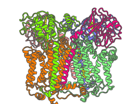 |
Experimental method | ELECTRON MICROSCOPY |
|---|---|---|
| Structural data | available | |
| Non-standard amino acids | None | |
| Missing residues/ Structural gap | Exists / Exists | |
| Resolution | 2.41 Å | |
| Deposition date | 2026-04-14 |
| pdb_00002a3f | Crystal structure of nitrophorin 2 aqua complex | ||
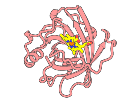 |
Experimental method | X-RAY DIFFRACTION |
|---|---|---|
| Structural data | available | |
| Non-standard amino acids | None | |
| Missing residues/ Structural gap | None / None | |
| Resolution | 1.4 Å | |
| Deposition date | 2005-06-24 |
| pdb_00002a3m | Structure of Desulfovibrio desulfuricans G20 tetraheme cytochrome (oxidized form) | ||
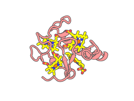 |
Experimental method | X-RAY DIFFRACTION |
|---|---|---|
| Structural data | available | |
| Non-standard amino acids | None | |
| Missing residues/ Structural gap | None / None | |
| Resolution | 1.5 Å | |
| Deposition date | 2005-06-25 |