
Number of data: 3514
Dataset: (without mutations)
| pdb_00002cn4 | The crystal structure of the secreted dimeric form of the hemophore HasA reveals a domain swapping with an exchanged heme ligand | ||
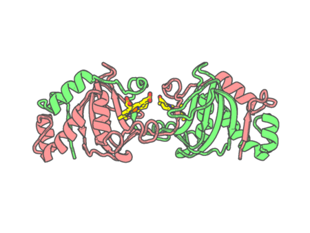 |
Experimental method | X-RAY DIFFRACTION |
|---|---|---|
| Structural data | available | |
| Non-standard amino acids | None | |
| Missing residues/ Structural gap | None / None | |
| Resolution | 2.3 Å | |
| Deposition date | 2006-05-18 |
| pdb_00002cpo | CHLOROPEROXIDASE | ||
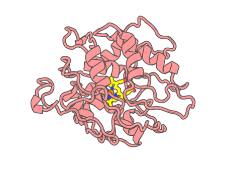 |
Experimental method | X-RAY DIFFRACTION |
|---|---|---|
| Structural data | available | |
| Non-standard amino acids | Included | |
| Missing residues/ Structural gap | None / None | |
| Resolution | 2.1 Å | |
| Deposition date | 1996-02-10 |
| pdb_00002cpp | HIGH-RESOLUTION CRYSTAL STRUCTURE OF CYTOCHROME P450-CAM | ||
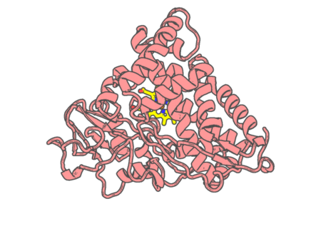 |
Experimental method | X-RAY DIFFRACTION |
|---|---|---|
| Structural data | available | |
| Non-standard amino acids | None | |
| Missing residues/ Structural gap | None / None | |
| Resolution | 1.63 Å | |
| Deposition date | 1987-04-06 |
| pdb_00002cth | CYTOCHROME C3 FROM DESULFOVIBRIO VULGARIS HILDENBOROUGH | ||
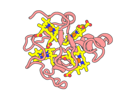 |
Experimental method | X-RAY DIFFRACTION |
|---|---|---|
| Structural data | available | |
| Non-standard amino acids | None | |
| Missing residues/ Structural gap | None / None | |
| Resolution | 1.67 Å | |
| Deposition date | 1997-06-18 |
| pdb_00002cvc | Crystal structure of High-Molecular Weight Cytochrome c from Desulfovibrio vulgaris (Hildenborough) | ||
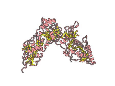 |
Experimental method | X-RAY DIFFRACTION |
|---|---|---|
| Structural data | available | |
| Non-standard amino acids | None | |
| Missing residues/ Structural gap | Exists / Exists | |
| Resolution | 2 Å | |
| Deposition date | 2005-06-02 |
| pdb_00002cxb | CRYSTALLIZATION AND X-RAY STRUCTURE DETERMINATION OF CYTOCHROME C2 FROM RHODOBACTER SPHAEROIDES IN THREE CRYSTAL FORMS | ||
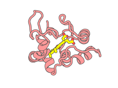 |
Experimental method | X-RAY DIFFRACTION |
|---|---|---|
| Structural data | available | |
| Non-standard amino acids | None | |
| Missing residues/ Structural gap | None / None | |
| Resolution | 1.95 Å | |
| Deposition date | 1994-04-21 |
| pdb_00002cy3 | CRYSTAL STRUCTURE OF CYTOCHROME C3 FROM DESULFOVIBRIO DESULFURICANS NORWAY AT 1.7 ANGSTROMS RESOLUTION | ||
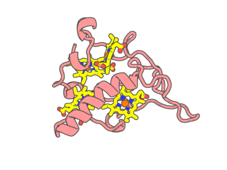 |
Experimental method | X-RAY DIFFRACTION |
|---|---|---|
| Structural data | available | |
| Non-standard amino acids | None | |
| Missing residues/ Structural gap | None / None | |
| Resolution | 1.7 Å | |
| Deposition date | 1994-07-01 |
| pdb_00002cym | EFFECTS OF AMINO ACID SUBSTITUTION ON THREE-DIMENSIONAL STRUCTURE: AN X-RAY ANALYSIS OF CYTOCHROME C3 FROM DESULFOVIBRIO VULGARIS HILDENBOROUGH AT 2 ANGSTROMS RESOLUTION | ||
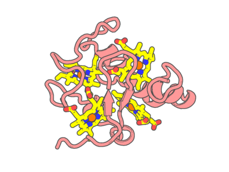 |
Experimental method | X-RAY DIFFRACTION |
|---|---|---|
| Structural data | available | |
| Non-standard amino acids | None | |
| Missing residues/ Structural gap | None / None | |
| Resolution | 2 Å | |
| Deposition date | 1993-09-29 |
| pdb_00002cyp | Crystal structure of yeast cytochrome C peroxidase refined at 1.7-angstroms resolution | ||
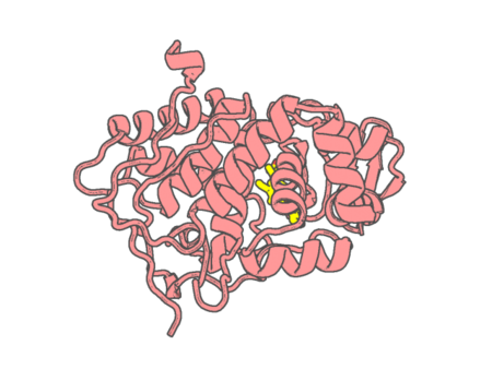 |
Experimental method | X-RAY DIFFRACTION |
|---|---|---|
| Structural data | available | |
| Non-standard amino acids | None | |
| Missing residues/ Structural gap | None / None | |
| Resolution | 1.7 Å | |
| Deposition date | 1985-08-27 |
| pdb_00002d09 | A Role for Active Site Water Molecules and Hydroxyl Groups of Substrate for Oxygen Activation in Cytochrome P450 158A2 | ||
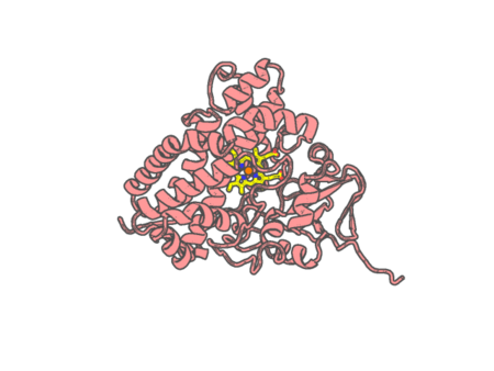 |
Experimental method | X-RAY DIFFRACTION |
|---|---|---|
| Structural data | available | |
| Non-standard amino acids | None | |
| Missing residues/ Structural gap | None / None | |
| Resolution | 1.8 Å | |
| Deposition date | 2005-07-30 |
| pdb_00002d0e | Substrate assited in Oxygen Activation in Cytochrome P450 158A2 | ||
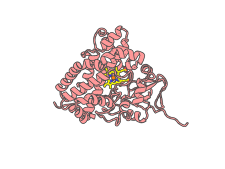 |
Experimental method | X-RAY DIFFRACTION |
|---|---|---|
| Structural data | available | |
| Non-standard amino acids | None | |
| Missing residues/ Structural gap | None / None | |
| Resolution | 2.15 Å | |
| Deposition date | 2005-08-02 |
| pdb_00002d0s | Crystal structure of the Cytochrome C552 from moderate thermophilic bacterium, hydrogenophilus thermoluteolus | ||
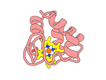 |
Experimental method | X-RAY DIFFRACTION |
|---|---|---|
| Structural data | available | |
| Non-standard amino acids | None | |
| Missing residues/ Structural gap | None / None | |
| Resolution | 2.2 Å | |
| Deposition date | 2005-08-08 |
| pdb_00002d0t | Crystal structure of 4-phenylimidazole bound form of human indoleamine 2,3-dioxygenase | ||
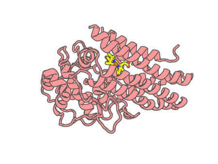 |
Experimental method | X-RAY DIFFRACTION |
|---|---|---|
| Structural data | available | |
| Non-standard amino acids | None | |
| Missing residues/ Structural gap | Exists / Exists | |
| Resolution | 2.3 Å | |
| Deposition date | 2005-08-08 |
| pdb_00002d0u | Crystal structure of cyanide bound form of human indoleamine 2,3-dioxygenase | ||
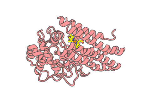 |
Experimental method | X-RAY DIFFRACTION |
|---|---|---|
| Structural data | available | |
| Non-standard amino acids | None | |
| Missing residues/ Structural gap | Exists / Exists | |
| Resolution | 3.4 Å | |
| Deposition date | 2005-08-08 |
| pdb_00002d0w | Crystal structure of cytochrome cL from Hyphomicrobium denitrificans | ||
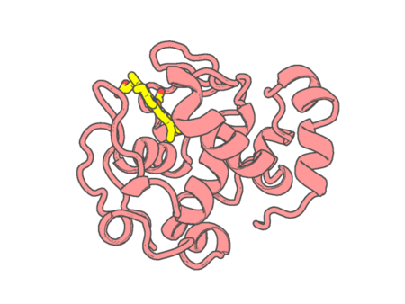 |
Experimental method | X-RAY DIFFRACTION |
|---|---|---|
| Structural data | available | |
| Non-standard amino acids | None | |
| Missing residues/ Structural gap | None / None | |
| Resolution | 1.98 Å | |
| Deposition date | 2005-08-10 |
| pdb_00002d2m | Structure of an extracellular giant hemoglobin of the gutless beard worm Oligobrachia mashikoi | ||
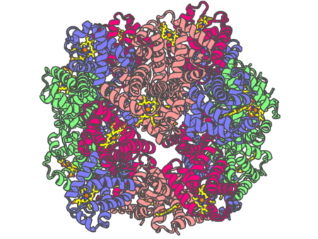 |
Experimental method | X-RAY DIFFRACTION |
|---|---|---|
| Structural data | available | |
| Non-standard amino acids | None | |
| Missing residues/ Structural gap | None / None | |
| Resolution | 2.85 Å | |
| Deposition date | 2005-09-12 |
| pdb_00002d2n | Structure of an extracellular giant hemoglobin of the gutless beard worm Oligobrachia mashikoi | ||
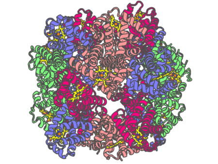 |
Experimental method | X-RAY DIFFRACTION |
|---|---|---|
| Structural data | available | |
| Non-standard amino acids | None | |
| Missing residues/ Structural gap | None / None | |
| Resolution | 3.2 Å | |
| Deposition date | 2005-09-12 |
| pdb_00002d3q | Crystal Structure of a Decolorizing Peroxidase (DyP) That Catalyses the Biological Oxidation of Anthraquinone Derivatives | ||
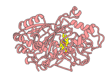 |
Experimental method | X-RAY DIFFRACTION |
|---|---|---|
| Structural data | available | |
| Non-standard amino acids | None | |
| Missing residues/ Structural gap | None / None | |
| Resolution | 2.8 Å | |
| Deposition date | 2005-09-30 |
| pdb_00002d5x | Crystal structure of carbonmonoxy horse hemoglobin complexed with L35 | ||
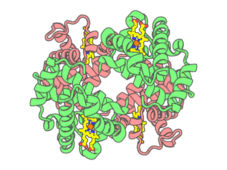 |
Experimental method | X-RAY DIFFRACTION |
|---|---|---|
| Structural data | available | |
| Non-standard amino acids | None | |
| Missing residues/ Structural gap | None / None | |
| Resolution | 1.45 Å | |
| Deposition date | 2005-11-08 |
| pdb_00002d5z | Crystal structure of T-state human hemoglobin complexed with three L35 molecules | ||
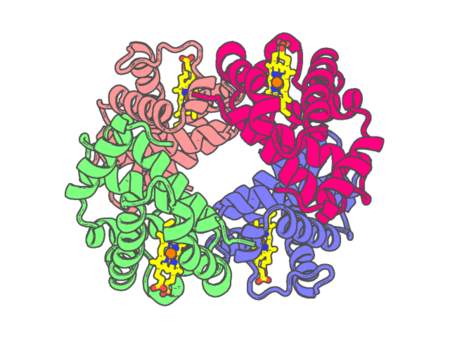 |
Experimental method | X-RAY DIFFRACTION |
|---|---|---|
| Structural data | available | |
| Non-standard amino acids | None | |
| Missing residues/ Structural gap | None / None | |
| Resolution | 1.45 Å | |
| Deposition date | 2005-11-08 |