
Number of data: 7652
Dataset: all data
| pdb_00009u7x | Crystal structure of the CYP105D18 mutant F191A from Streptomyces laurentii | ||
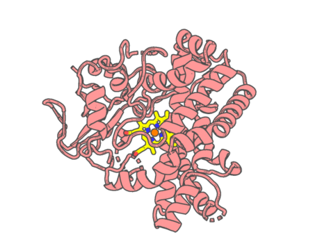 |
Experimental method | X-RAY DIFFRACTION |
|---|---|---|
| Structural data | available | |
| Non-standard amino acids | None | |
| Missing residues/ Structural gap | Exists / Exists | |
| Resolution | 1.45 Å | |
| Deposition date | 2025-03-25 |
| pdb_00009ub3 | The structure of the apo-AglA from Streptomyces monomycini | ||
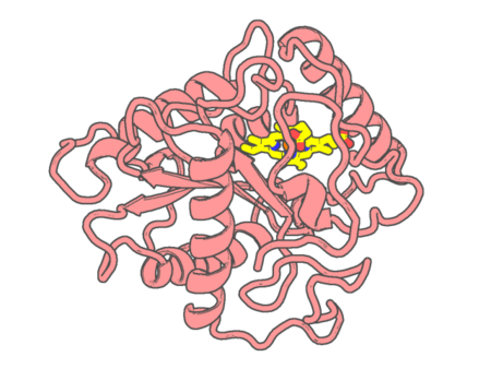 |
Experimental method | X-RAY DIFFRACTION |
|---|---|---|
| Structural data | available | |
| Non-standard amino acids | None | |
| Missing residues/ Structural gap | None / None | |
| Resolution | 2.06 Å | |
| Deposition date | 2025-04-02 |
| pdb_00009ub5 | The structure of the AglA-Arg complex | ||
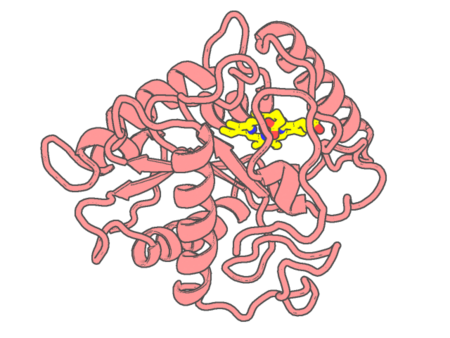 |
Experimental method | X-RAY DIFFRACTION |
|---|---|---|
| Structural data | available | |
| Non-standard amino acids | None | |
| Missing residues/ Structural gap | None / None | |
| Resolution | 1.83 Å | |
| Deposition date | 2025-04-02 |
| pdb_00009uba | The structure of the AglA_K196R-Arg complex | ||
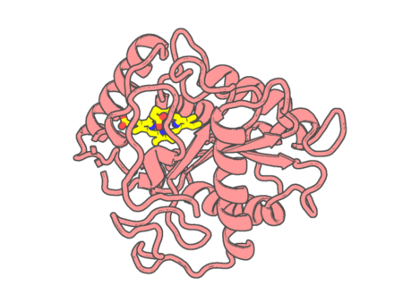 |
Experimental method | X-RAY DIFFRACTION |
|---|---|---|
| Structural data | available | |
| Non-standard amino acids | None | |
| Missing residues/ Structural gap | Exists / Exists | |
| Resolution | 1.47 Å | |
| Deposition date | 2025-04-02 |
| pdb_00009ubb | The structure of the AglA_K225E-Arg complex | ||
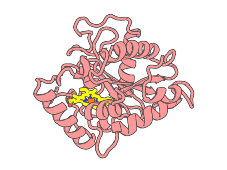 |
Experimental method | X-RAY DIFFRACTION |
|---|---|---|
| Structural data | available | |
| Non-standard amino acids | None | |
| Missing residues/ Structural gap | Exists / Exists | |
| Resolution | 1.41 Å | |
| Deposition date | 2025-04-02 |
| pdb_00009ubs | The structure of the AglA_T220R-Arg complex | ||
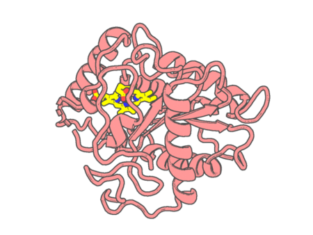 |
Experimental method | X-RAY DIFFRACTION |
|---|---|---|
| Structural data | available | |
| Non-standard amino acids | None | |
| Missing residues/ Structural gap | Exists / Exists | |
| Resolution | 1.49 Å | |
| Deposition date | 2025-04-03 |
| pdb_00009ubt | The structure of the AglA-Ampn-Arg complex | ||
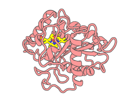 |
Experimental method | X-RAY DIFFRACTION |
|---|---|---|
| Structural data | available | |
| Non-standard amino acids | None | |
| Missing residues/ Structural gap | None / None | |
| Resolution | 1.596 Å | |
| Deposition date | 2025-04-03 |
| pdb_00009ufb | Ubiquinol Binding Site of Cytochrome bo3 from A.b | ||
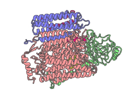 |
Experimental method | ELECTRON MICROSCOPY |
|---|---|---|
| Structural data | available | |
| Non-standard amino acids | None | |
| Missing residues/ Structural gap | None / None | |
| Resolution | 3.05 Å | |
| Deposition date | 2025-04-10 |
| pdb_00009uft | Ubiquinol Binding Site of Cytochrome bo3 from Acinetobacter baumannii | ||
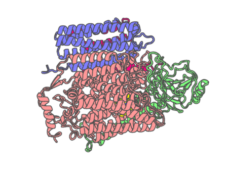 |
Experimental method | ELECTRON MICROSCOPY |
|---|---|---|
| Structural data | available | |
| Non-standard amino acids | None | |
| Missing residues/ Structural gap | None / None | |
| Resolution | None | |
| Deposition date | 2025-04-10 |
| pdb_00009ufv | Ubiquinol Binding Site of Cytochrome bo3 from Acinetobacter baumannii | ||
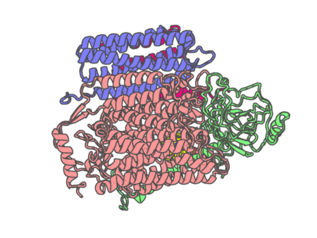 |
Experimental method | ELECTRON MICROSCOPY |
|---|---|---|
| Structural data | available | |
| Non-standard amino acids | None | |
| Missing residues/ Structural gap | None / None | |
| Resolution | 3.56 Å | |
| Deposition date | 2025-04-10 |
| pdb_00009ufw | Ubiquinol Binding Site of Cytochrome bo3 from Acinetobacter baumannii | ||
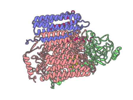 |
Experimental method | ELECTRON MICROSCOPY |
|---|---|---|
| Structural data | available | |
| Non-standard amino acids | None | |
| Missing residues/ Structural gap | None / None | |
| Resolution | 3.12 Å | |
| Deposition date | 2025-04-10 |
| pdb_00009ufz | Ubiquinol Binding Site of Cytochrome bo3 from Acinetobacter baumannii | ||
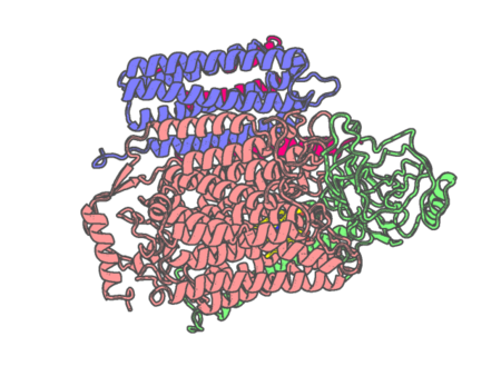 |
Experimental method | ELECTRON MICROSCOPY |
|---|---|---|
| Structural data | available | |
| Non-standard amino acids | None | |
| Missing residues/ Structural gap | None / None | |
| Resolution | 3.28 Å | |
| Deposition date | 2025-04-10 |
| pdb_00009ule | Crystal structure of CYP105A1 R84A complexed with diclofenac (DIF) at room temperature | ||
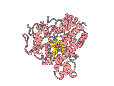 |
Experimental method | X-RAY DIFFRACTION |
|---|---|---|
| Structural data | available | |
| Non-standard amino acids | None | |
| Missing residues/ Structural gap | None / None | |
| Resolution | 1.86 Å | |
| Deposition date | 2025-04-19 |
| pdb_00009ulw | Crystal Structure of Cytochrome P450 GpeC | ||
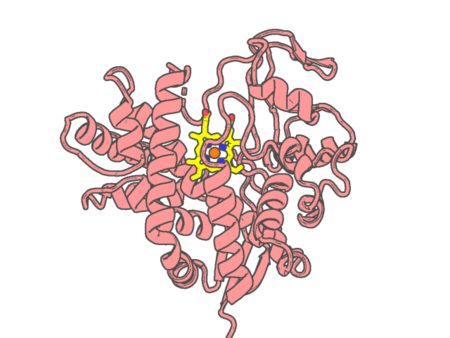 |
Experimental method | X-RAY DIFFRACTION |
|---|---|---|
| Structural data | available | |
| Non-standard amino acids | None | |
| Missing residues/ Structural gap | Exists / Exists | |
| Resolution | 2.62 Å | |
| Deposition date | 2025-04-21 |
| pdb_00009uxz | Crystal structure of the P450BM3 triple mutant F87A/L215W/H408W from Priestia megaterium | ||
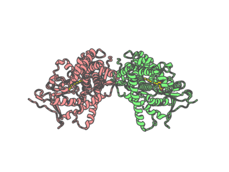 |
Experimental method | X-RAY DIFFRACTION |
|---|---|---|
| Structural data | available | |
| Non-standard amino acids | None | |
| Missing residues/ Structural gap | None / None | |
| Resolution | 1.7 Å | |
| Deposition date | 2025-05-14 |
| pdb_00009uy0 | Crystal structure of the CYP153A double mutant L354I/V456G from Marinobacter aquaeolei | ||
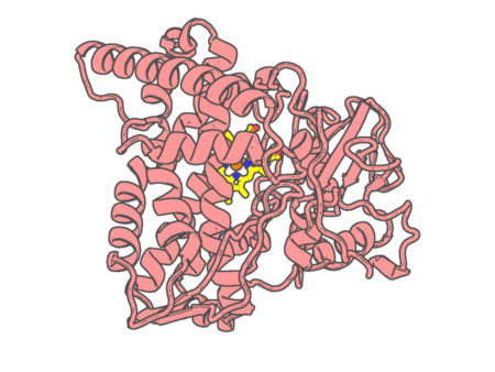 |
Experimental method | X-RAY DIFFRACTION |
|---|---|---|
| Structural data | available | |
| Non-standard amino acids | None | |
| Missing residues/ Structural gap | None / None | |
| Resolution | 1.96 Å | |
| Deposition date | 2025-05-14 |
| pdb_00009uyc | TxtE mutant-H176FA248F With trp | ||
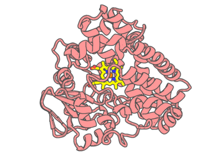 |
Experimental method | X-RAY DIFFRACTION |
|---|---|---|
| Structural data | available | |
| Non-standard amino acids | None | |
| Missing residues/ Structural gap | None / None | |
| Resolution | 1.53 Å | |
| Deposition date | 2025-05-15 |
| pdb_00009uyy | Crystal structure of the indoleamine 2,3-dioxygenagse 2 (IDO2) complexed with L-Trp | ||
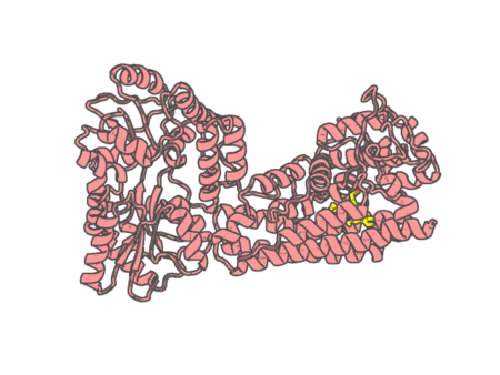 |
Experimental method | X-RAY DIFFRACTION |
|---|---|---|
| Structural data | available | |
| Non-standard amino acids | None | |
| Missing residues/ Structural gap | Exists / Exists | |
| Resolution | 2.25 Å | |
| Deposition date | 2025-05-16 |
| pdb_00009uyz | Crystal structure of the indoleamine 2,3-dioxygenagse 2 (IDO2) complexed with 5-HT | ||
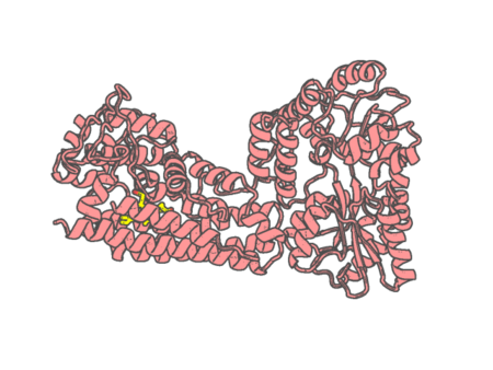 |
Experimental method | X-RAY DIFFRACTION |
|---|---|---|
| Structural data | unavailable | |
| Non-standard amino acids | None | |
| Missing residues/ Structural gap | Exists / Exists | |
| Resolution | 2.45 Å | |
| Deposition date | 2025-05-16 |
| pdb_00009uz0 | Crystal structure of the indoleamine 2,3-dioxygenagse 2 (IDO2) as resting state | ||
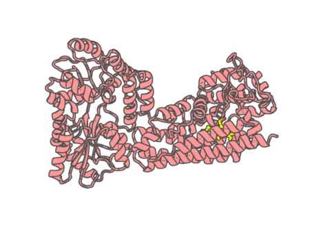 |
Experimental method | X-RAY DIFFRACTION |
|---|---|---|
| Structural data | available | |
| Non-standard amino acids | None | |
| Missing residues/ Structural gap | Exists / Exists | |
| Resolution | 2.45 Å | |
| Deposition date | 2025-05-16 |