
Number of data: 3898
Dataset: (exptl_method: X-ray) and (resolution_limit=2.0)
| pdb_00002fal | X-RAY CRYSTAL STRUCTURE OF FERRIC APLYSIA LIMACINA MYOGLOBIN IN DIFFERENT LIGANDED STATES | ||
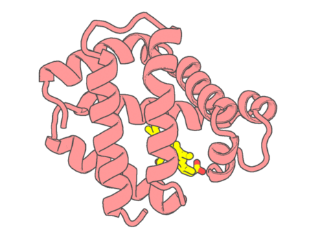 |
Experimental method | X-RAY DIFFRACTION |
|---|---|---|
| Structural data | available | |
| Non-standard amino acids | None | |
| Missing residues/ Structural gap | None / None | |
| Resolution | 1.8 Å | |
| Deposition date | 1993-06-14 |
| pdb_00002fam | X-RAY CRYSTAL STRUCTURE OF FERRIC APLYSIA LIMACINA MYOGLOBIN IN DIFFERENT LIGANDED STATES | ||
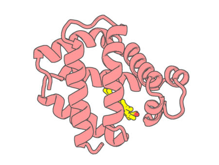 |
Experimental method | X-RAY DIFFRACTION |
|---|---|---|
| Structural data | available | |
| Non-standard amino acids | None | |
| Missing residues/ Structural gap | None / None | |
| Resolution | 2 Å | |
| Deposition date | 1993-07-20 |
| pdb_00002fc1 | Heme NO Complex in NOS | ||
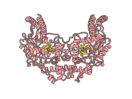 |
Experimental method | X-RAY DIFFRACTION |
|---|---|---|
| Structural data | available | |
| Non-standard amino acids | None | |
| Missing residues/ Structural gap | Exists / None | |
| Resolution | 2 Å | |
| Deposition date | 2005-12-10 |
| pdb_00002fdu | Microsomal P450 2A6 with the inhibitor N,N-Dimethyl(5-(pyridin-3-yl)furan-2-yl)methanamine bound | ||
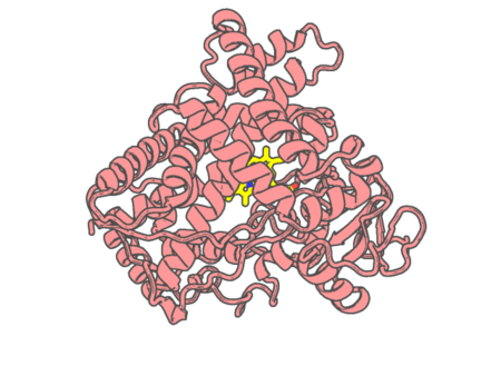 |
Experimental method | X-RAY DIFFRACTION |
|---|---|---|
| Structural data | available | |
| Non-standard amino acids | None | |
| Missing residues/ Structural gap | None / None | |
| Resolution | 1.85 Å | |
| Deposition date | 2005-12-14 |
| pdb_00002fdv | Microsomal P450 2A6 with the inhibitor N-Methyl(5-(pyridin-3-yl)furan-2-yl)methanamine bound | ||
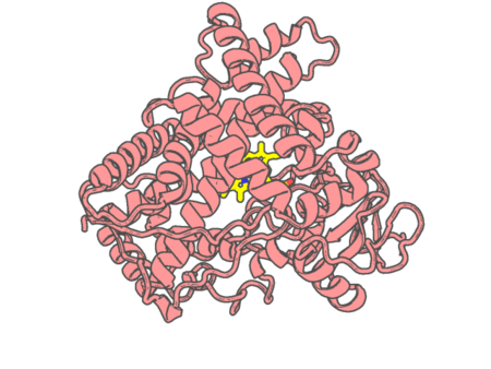 |
Experimental method | X-RAY DIFFRACTION |
|---|---|---|
| Structural data | available | |
| Non-standard amino acids | None | |
| Missing residues/ Structural gap | None / None | |
| Resolution | 1.65 Å | |
| Deposition date | 2005-12-14 |
| pdb_00002fdy | Microsomal P450 2A6 with the inhibitor Adrithiol bound | ||
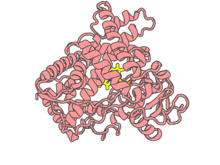 |
Experimental method | X-RAY DIFFRACTION |
|---|---|---|
| Structural data | available | |
| Non-standard amino acids | None | |
| Missing residues/ Structural gap | None / None | |
| Resolution | 1.95 Å | |
| Deposition date | 2005-12-14 |
| pdb_00002ffn | The E41Q mutant of tetraheme cytochrome c3 from Desulfovibrio Vulgaris Miyazaki F | ||
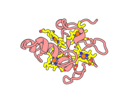 |
Experimental method | X-RAY DIFFRACTION |
|---|---|---|
| Structural data | available | |
| Non-standard amino acids | None | |
| Missing residues/ Structural gap | None / None | |
| Resolution | 1.8 Å | |
| Deposition date | 2005-12-20 |
| pdb_00002fkz | Reduced (All Ferrous) form of the Azotobacter vinelandii bacterioferritin | ||
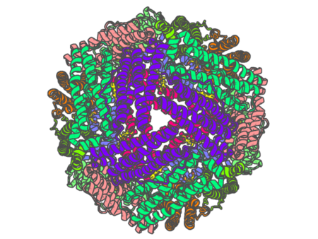 |
Experimental method | X-RAY DIFFRACTION |
|---|---|---|
| Structural data | available | |
| Non-standard amino acids | None | |
| Missing residues/ Structural gap | None / None | |
| Resolution | 2 Å | |
| Deposition date | 2006-01-05 |
| pdb_00002frf | Horse Heart Myoglobin, Nitrite Adduct, Crystal Soak | ||
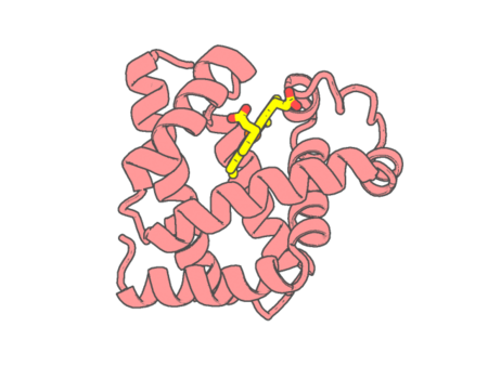 |
Experimental method | X-RAY DIFFRACTION |
|---|---|---|
| Structural data | available | |
| Non-standard amino acids | None | |
| Missing residues/ Structural gap | None / None | |
| Resolution | 1.2 Å | |
| Deposition date | 2006-01-19 |
| pdb_00002fri | Horse Heart Myoglobin, Nitrite Adduct, Co-crystallized | ||
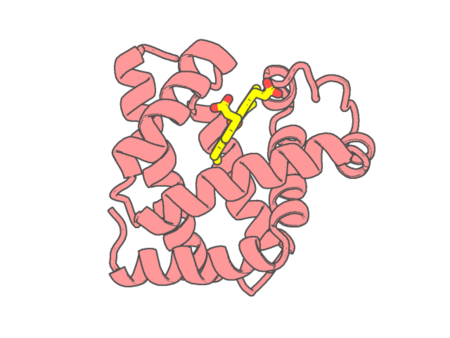 |
Experimental method | X-RAY DIFFRACTION |
|---|---|---|
| Structural data | available | |
| Non-standard amino acids | None | |
| Missing residues/ Structural gap | None / None | |
| Resolution | 1.6 Å | |
| Deposition date | 2006-01-19 |
| pdb_00002frj | Nitrosyl Horse Heart Myoglobin, Nitrite/Dithionite Method | ||
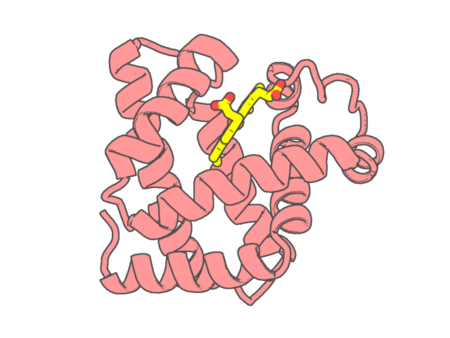 |
Experimental method | X-RAY DIFFRACTION |
|---|---|---|
| Structural data | available | |
| Non-standard amino acids | None | |
| Missing residues/ Structural gap | None / None | |
| Resolution | 1.3 Å | |
| Deposition date | 2006-01-19 |
| pdb_00002frk | Nitrosyl Horse Heart Myoglobin, Nitric Oxide Gas Method | ||
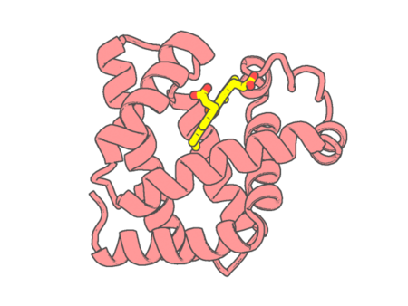 |
Experimental method | X-RAY DIFFRACTION |
|---|---|---|
| Structural data | available | |
| Non-standard amino acids | None | |
| Missing residues/ Structural gap | None / None | |
| Resolution | 1.3 Å | |
| Deposition date | 2006-01-19 |
| pdb_00002fw5 | Diheme cytochrome c from Rhodobacter sphaeroides | ||
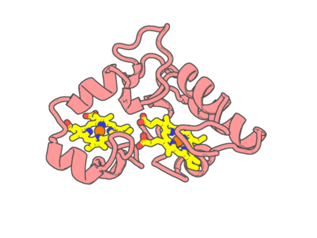 |
Experimental method | X-RAY DIFFRACTION |
|---|---|---|
| Structural data | available | |
| Non-standard amino acids | None | |
| Missing residues/ Structural gap | None / None | |
| Resolution | 2 Å | |
| Deposition date | 2006-02-01 |
| pdb_00002fwt | Crystal structure of DHC purified from Rhodobacter sphaeroides | ||
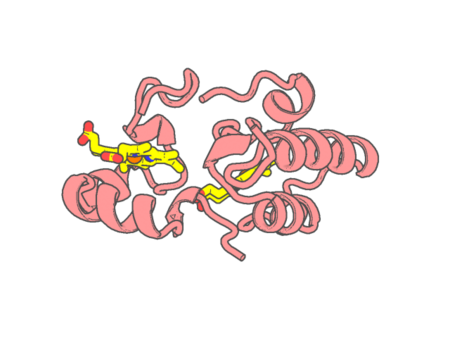 |
Experimental method | X-RAY DIFFRACTION |
|---|---|---|
| Structural data | available | |
| Non-standard amino acids | None | |
| Missing residues/ Structural gap | Exists / Exists | |
| Resolution | 1.85 Å | |
| Deposition date | 2006-02-03 |
| pdb_00002g0r | Unphotolyzed CO-bound L29F Myoglobin | ||
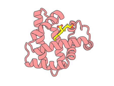 |
Experimental method | X-RAY DIFFRACTION |
|---|---|---|
| Structural data | available | |
| Non-standard amino acids | None | |
| Missing residues/ Structural gap | None / None | |
| Resolution | 1.95 Å | |
| Deposition date | 2006-02-13 |
| pdb_00002g0s | Unphotolyzed CO-bound L29F Myoglobin, crystal 2 | ||
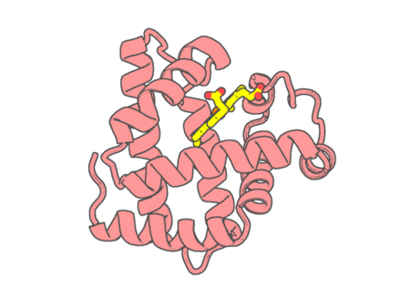 |
Experimental method | X-RAY DIFFRACTION |
|---|---|---|
| Structural data | available | |
| Non-standard amino acids | None | |
| Missing residues/ Structural gap | None / None | |
| Resolution | 1.9 Å | |
| Deposition date | 2006-02-13 |
| pdb_00002g0v | Photolyzed CO L29F Myoglobin: 100ps | ||
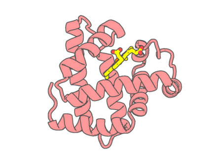 |
Experimental method | X-RAY DIFFRACTION |
|---|---|---|
| Structural data | available | |
| Non-standard amino acids | None | |
| Missing residues/ Structural gap | None / None | |
| Resolution | 1.95 Å | |
| Deposition date | 2006-02-13 |
| pdb_00002g0x | Photolyzed CO L29F Myoglobin: 316ps | ||
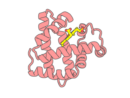 |
Experimental method | X-RAY DIFFRACTION |
|---|---|---|
| Structural data | available | |
| Non-standard amino acids | None | |
| Missing residues/ Structural gap | None / None | |
| Resolution | 1.95 Å | |
| Deposition date | 2006-02-13 |
| pdb_00002g0z | Photolyzed CO L29F Myoglobin: 1ns | ||
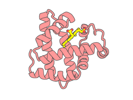 |
Experimental method | X-RAY DIFFRACTION |
|---|---|---|
| Structural data | available | |
| Non-standard amino acids | None | |
| Missing residues/ Structural gap | None / None | |
| Resolution | 1.95 Å | |
| Deposition date | 2006-02-13 |
| pdb_00002g10 | Photolyzed CO L29F Myoglobin: 3.16ns | ||
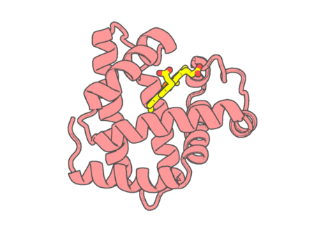 |
Experimental method | X-RAY DIFFRACTION |
|---|---|---|
| Structural data | available | |
| Non-standard amino acids | None | |
| Missing residues/ Structural gap | None / None | |
| Resolution | 1.9 Å | |
| Deposition date | 2006-02-13 |