
Number of data: 7660
Dataset: all data
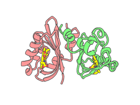
| pdb_00001s67 | Crystal structure of heme domain of direct oxygen sensor from E. coli | ||
 |
Experimental method | X-RAY DIFFRACTION |
|---|---|---|
| Structural data | available | |
| Non-standard amino acids | None | |
| Missing residues/ Structural gap | None / None | |
| Resolution | 1.5 Å | |
| Deposition date | 2004-01-22 |
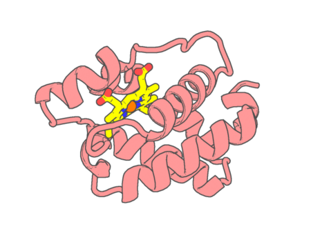
| pdb_00001s69 | The X-ray structure of the cyanobacteria Synechocystis hemoglobin "cyanoglobin" with cyanide ligand | ||
 |
Experimental method | X-RAY DIFFRACTION |
|---|---|---|
| Structural data | available | |
| Non-standard amino acids | None | |
| Missing residues/ Structural gap | None / None | |
| Resolution | 1.68 Å | |
| Deposition date | 2004-01-22 |
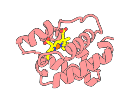
| pdb_00001s6a | The X-ray structure of the cyanobacteria Synechocystis hemoglobin "cyanoglobin" with azide ligand | ||
 |
Experimental method | X-RAY DIFFRACTION |
|---|---|---|
| Structural data | available | |
| Non-standard amino acids | None | |
| Missing residues/ Structural gap | None / None | |
| Resolution | 1.69 Å | |
| Deposition date | 2004-01-22 |
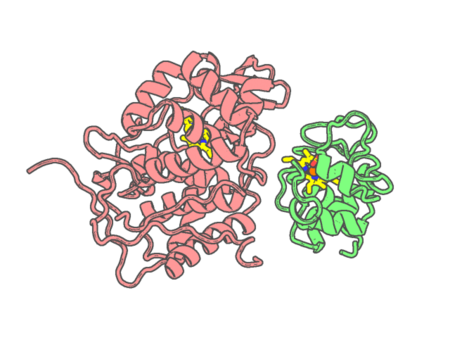
| pdb_00001s6v | Structure of a cytochrome c peroxidase-cytochrome c site specific cross-link | ||
 |
Experimental method | X-RAY DIFFRACTION |
|---|---|---|
| Structural data | available | |
| Non-standard amino acids | None | |
| Missing residues/ Structural gap | None / None | |
| Resolution | 1.88 Å | |
| Deposition date | 2004-01-27 |
| pdb_00001sch | PEANUT PEROXIDASE | ||
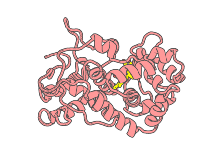 |
Experimental method | X-RAY DIFFRACTION |
|---|---|---|
| Structural data | available | |
| Non-standard amino acids | Included | |
| Missing residues/ Structural gap | None / None | |
| Resolution | 2.56 Å | |
| Deposition date | 1996-01-23 |
| pdb_00001sdk | CROSS-LINKED, CARBONMONOXY HEMOGLOBIN A | ||
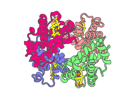 |
Experimental method | X-RAY DIFFRACTION |
|---|---|---|
| Structural data | available | |
| Non-standard amino acids | None | |
| Missing residues/ Structural gap | None / None | |
| Resolution | 1.8 Å | |
| Deposition date | 1996-02-26 |
| pdb_00001sdl | CROSS-LINKED, CARBONMONOXY HEMOGLOBIN A | ||
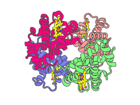 |
Experimental method | X-RAY DIFFRACTION |
|---|---|---|
| Structural data | available | |
| Non-standard amino acids | None | |
| Missing residues/ Structural gap | None / None | |
| Resolution | 1.8 Å | |
| Deposition date | 1996-02-26 |
| pdb_00001se6 | Crystal Structure of Streptomyces Coelicolor A3(2) CYP158A2 from antibiotic biosynthetic pathways | ||
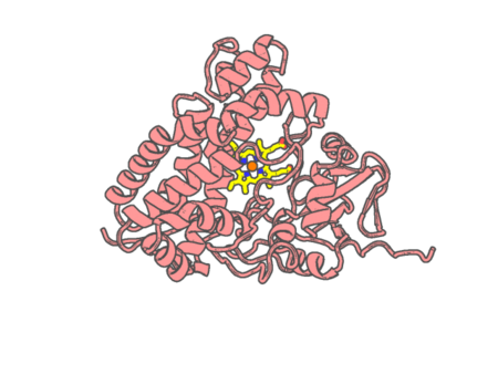 |
Experimental method | X-RAY DIFFRACTION |
|---|---|---|
| Structural data | available | |
| Non-standard amino acids | None | |
| Missing residues/ Structural gap | None / None | |
| Resolution | 1.75 Å | |
| Deposition date | 2004-02-16 |
| pdb_00001sh4 | Solution structure of oxidized bovine microsomal cytochrome B5 Mutant V45H | ||
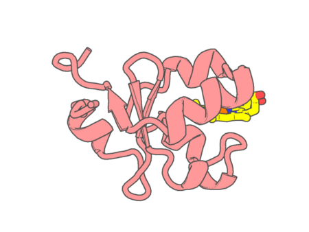 |
Experimental method | SOLUTION NMR |
|---|---|---|
| Structural data | available | |
| Non-standard amino acids | None | |
| Missing residues/ Structural gap | Exists / Exists | |
| Resolution | None | |
| Deposition date | 2004-02-25 |
| pdb_00001shr | Crystal structure of ferrocyanide bound human hemoglobin A2 at 1.88A resolution | ||
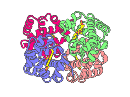 |
Experimental method | X-RAY DIFFRACTION |
|---|---|---|
| Structural data | available | |
| Non-standard amino acids | None | |
| Missing residues/ Structural gap | None / None | |
| Resolution | 1.88 Å | |
| Deposition date | 2004-02-26 |
| pdb_00001si4 | Crystal structure of Human hemoglobin A2 (in R2 state) at 2.2 A resolution | ||
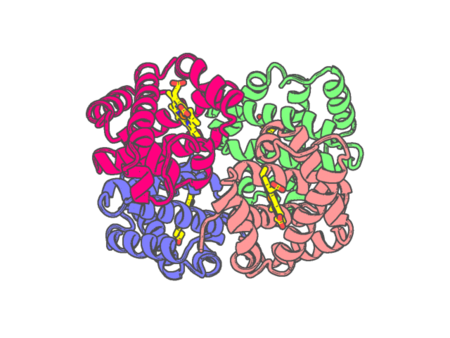 |
Experimental method | X-RAY DIFFRACTION |
|---|---|---|
| Structural data | available | |
| Non-standard amino acids | None | |
| Missing residues/ Structural gap | None / None | |
| Resolution | 2.2 Å | |
| Deposition date | 2004-02-27 |
| pdb_00001si6 | Crystal structure of cimex nitrophorin complex with CO | ||
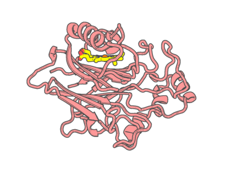 |
Experimental method | X-RAY DIFFRACTION |
|---|---|---|
| Structural data | available | |
| Non-standard amino acids | None | |
| Missing residues/ Structural gap | None / None | |
| Resolution | 1.45 Å | |
| Deposition date | 2004-02-27 |
| pdb_00001si8 | Crystal structure of E. faecalis catalase | ||
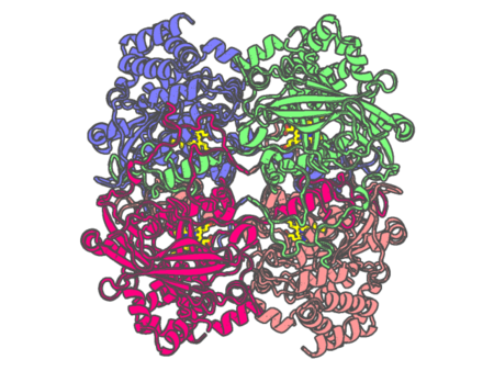 |
Experimental method | X-RAY DIFFRACTION |
|---|---|---|
| Structural data | available | |
| Non-standard amino acids | None | |
| Missing residues/ Structural gap | None / None | |
| Resolution | 2.3 Å | |
| Deposition date | 2004-02-28 |
| pdb_00001siw | Crystal structure of the apomolybdo-NarGHI | ||
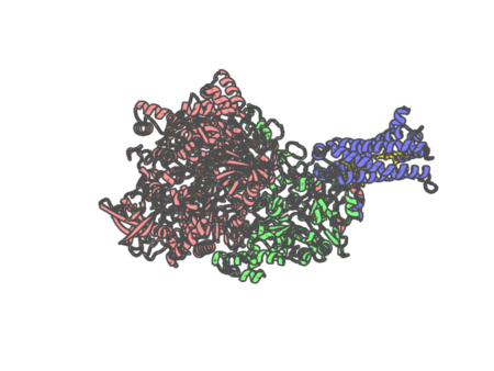 |
Experimental method | X-RAY DIFFRACTION |
|---|---|---|
| Structural data | available | |
| Non-standard amino acids | Included | |
| Missing residues/ Structural gap | Exists / Exists | |
| Resolution | 2.2 Å | |
| Deposition date | 2004-03-01 |
| pdb_00001sj2 | Crystal structure of Mycobacterium tuberculosis catalase-peroxidase | ||
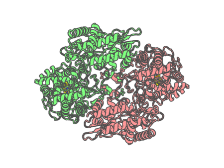 |
Experimental method | X-RAY DIFFRACTION |
|---|---|---|
| Structural data | available | |
| Non-standard amino acids | None | |
| Missing residues/ Structural gap | None / None | |
| Resolution | 2.41 Å | |
| Deposition date | 2004-03-02 |
| pdb_00001sk7 | Structural Basis for Novel Delta-Regioselective Heme Oxygenation in the Opportunistic Pathogen Pseudomonas aeruginosa | ||
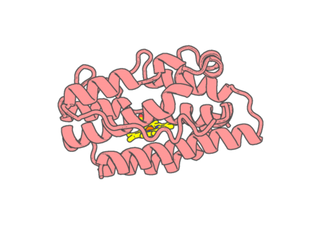 |
Experimental method | X-RAY DIFFRACTION |
|---|---|---|
| Structural data | available | |
| Non-standard amino acids | None | |
| Missing residues/ Structural gap | None / None | |
| Resolution | 1.6 Å | |
| Deposition date | 2004-03-04 |
| pdb_00001smi | A single mutation of P450 BM3 induces the conformational rearrangement seen upon substrate-binding in wild-type enzyme | ||
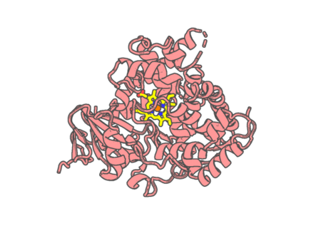 |
Experimental method | X-RAY DIFFRACTION |
|---|---|---|
| Structural data | available | |
| Non-standard amino acids | None | |
| Missing residues/ Structural gap | Exists / Exists | |
| Resolution | 2 Å | |
| Deposition date | 2004-03-09 |
| pdb_00001smj | Structure of the A264E mutant of cytochrome P450 BM3 complexed with palmitoleate | ||
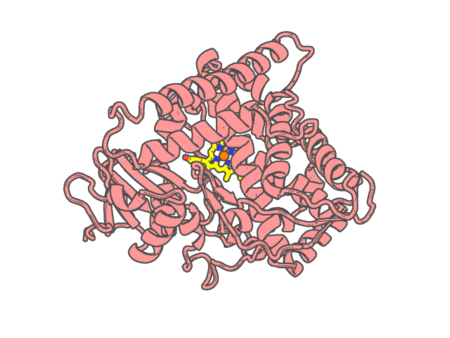 |
Experimental method | X-RAY DIFFRACTION |
|---|---|---|
| Structural data | available | |
| Non-standard amino acids | None | |
| Missing residues/ Structural gap | None / None | |
| Resolution | 2.75 Å | |
| Deposition date | 2004-03-09 |
| pdb_00001sof | Crystal structure of the azotobacter vinelandii bacterioferritin at 2.6 A resolution | ||
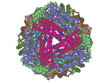 |
Experimental method | X-RAY DIFFRACTION |
|---|---|---|
| Structural data | available | |
| Non-standard amino acids | None | |
| Missing residues/ Structural gap | None / None | |
| Resolution | 2.6 Å | |
| Deposition date | 2004-03-14 |
| pdb_00001sog | Cyrstal Structure of Cytochrome c Peroxidase Mutant: CcPK2M2 | ||
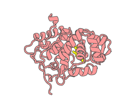 |
Experimental method | X-RAY DIFFRACTION |
|---|---|---|
| Structural data | available | |
| Non-standard amino acids | None | |
| Missing residues/ Structural gap | None / None | |
| Resolution | 1.85 Å | |
| Deposition date | 2004-03-14 |