
Number of data: 7660
Dataset: all data
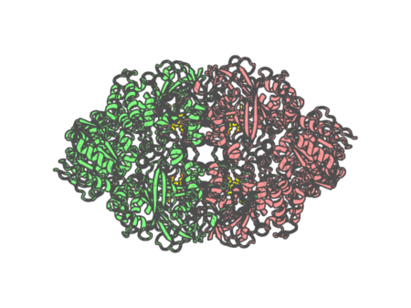
| pdb_00001sy7 | Crystal structure of the catalase-1 from Neurospora crassa, native structure at 1.75A resolution. | ||
 |
Experimental method | X-RAY DIFFRACTION |
|---|---|---|
| Structural data | available | |
| Non-standard amino acids | None | |
| Missing residues/ Structural gap | None / None | |
| Resolution | 1.75 Å | |
| Deposition date | 2004-04-01 |
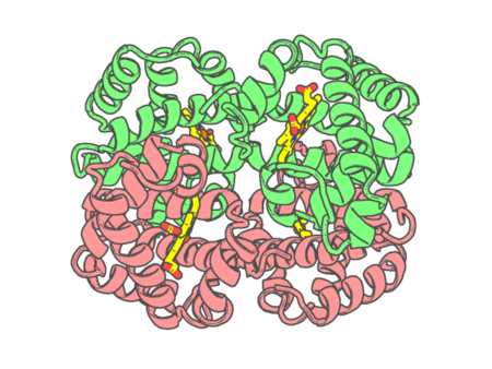
| pdb_00001t1n | CRYSTAL STRUCTURE OF CARBONMONOXY HEMOGLOBIN | ||
 |
Experimental method | X-RAY DIFFRACTION |
|---|---|---|
| Structural data | available | |
| Non-standard amino acids | None | |
| Missing residues/ Structural gap | None / None | |
| Resolution | 2.2 Å | |
| Deposition date | 1999-03-05 |
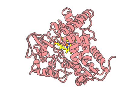
| pdb_00001t2b | Crystal Structure of cytochrome P450cin complexed with its substrate 1,8-cineole | ||
 |
Experimental method | X-RAY DIFFRACTION |
|---|---|---|
| Structural data | available | |
| Non-standard amino acids | None | |
| Missing residues/ Structural gap | None / None | |
| Resolution | 1.7 Å | |
| Deposition date | 2004-04-20 |
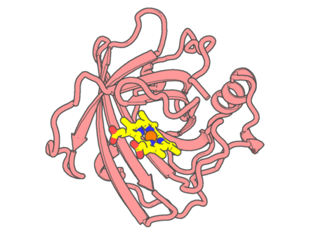
| pdb_00001t68 | Crystal structure of nitrophorin 2 complex with NO | ||
 |
Experimental method | X-RAY DIFFRACTION |
|---|---|---|
| Structural data | available | |
| Non-standard amino acids | None | |
| Missing residues/ Structural gap | None / None | |
| Resolution | 1.45 Å | |
| Deposition date | 2004-05-05 |
| pdb_00001t85 | Crystal Structure of the Ferrous CO-bound Cytochrome P450cam Mutant (L358P/C334A) | ||
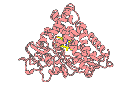 |
Experimental method | X-RAY DIFFRACTION |
|---|---|---|
| Structural data | available | |
| Non-standard amino acids | None | |
| Missing residues/ Structural gap | None / None | |
| Resolution | 1.8 Å | |
| Deposition date | 2004-05-11 |
| pdb_00001t86 | Crystal Structure of the Ferrous Cytochrome P450cam Mutant (L358P/C334A) | ||
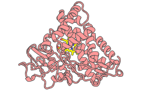 |
Experimental method | X-RAY DIFFRACTION |
|---|---|---|
| Structural data | available | |
| Non-standard amino acids | None | |
| Missing residues/ Structural gap | None / None | |
| Resolution | 1.9 Å | |
| Deposition date | 2004-05-11 |
| pdb_00001t87 | Crystal Structure of the Ferrous CO-bound Cytochrome P450cam (C334A) | ||
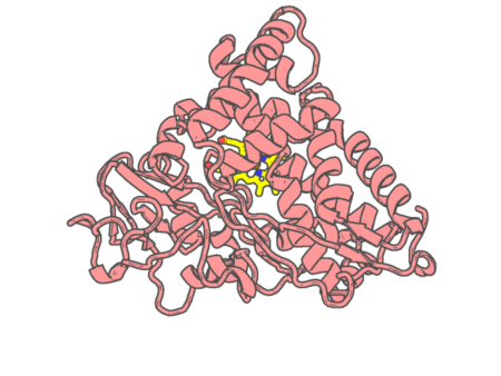 |
Experimental method | X-RAY DIFFRACTION |
|---|---|---|
| Structural data | available | |
| Non-standard amino acids | None | |
| Missing residues/ Structural gap | None / None | |
| Resolution | 1.8 Å | |
| Deposition date | 2004-05-11 |
| pdb_00001t88 | Crystal Structure of the Ferrous Cytochrome P450cam (C334A) | ||
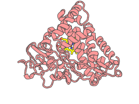 |
Experimental method | X-RAY DIFFRACTION |
|---|---|---|
| Structural data | available | |
| Non-standard amino acids | None | |
| Missing residues/ Structural gap | None / None | |
| Resolution | 1.9 Å | |
| Deposition date | 2004-05-11 |
| pdb_00001t93 | Evidence for Multiple Substrate Recognition and Molecular Mechanism of C-C reaction by Cytochrome P450 CYP158A2 from Streptomyces Coelicolor A3(2) | ||
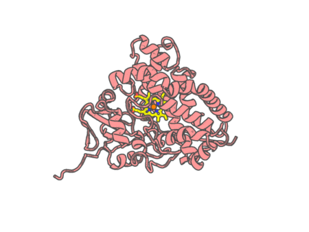 |
Experimental method | X-RAY DIFFRACTION |
|---|---|---|
| Structural data | available | |
| Non-standard amino acids | None | |
| Missing residues/ Structural gap | None / None | |
| Resolution | 1.62 Å | |
| Deposition date | 2004-05-14 |
| pdb_00001tes | OXYGEN BINDING MUSCLE PROTEIN | ||
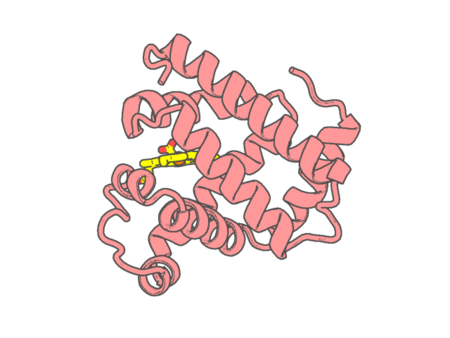 |
Experimental method | X-RAY DIFFRACTION |
|---|---|---|
| Structural data | available | |
| Non-standard amino acids | None | |
| Missing residues/ Structural gap | None / None | |
| Resolution | 1.7 Å | |
| Deposition date | 1996-05-06 |
| pdb_00001tgu | The crystal structure of bovine liver catalase without NADPH | ||
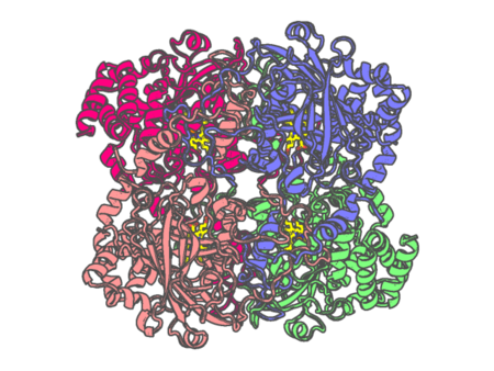 |
Experimental method | X-RAY DIFFRACTION |
|---|---|---|
| Structural data | available | |
| Non-standard amino acids | None | |
| Missing residues/ Structural gap | None / None | |
| Resolution | 2.8 Å | |
| Deposition date | 2004-05-31 |
| pdb_00001th2 | crystal structure of NADPH depleted bovine liver catalase complexed with azide | ||
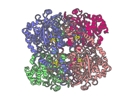 |
Experimental method | X-RAY DIFFRACTION |
|---|---|---|
| Structural data | available | |
| Non-standard amino acids | None | |
| Missing residues/ Structural gap | None / None | |
| Resolution | 2.8 Å | |
| Deposition date | 2004-06-01 |
| pdb_00001th3 | Crystal structure of NADPH depleted bovine live catalase complexed with cyanide | ||
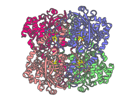 |
Experimental method | X-RAY DIFFRACTION |
|---|---|---|
| Structural data | available | |
| Non-standard amino acids | None | |
| Missing residues/ Structural gap | None / None | |
| Resolution | 2.8 Å | |
| Deposition date | 2004-06-01 |
| pdb_00001th4 | crystal structure of NADPH depleted bovine liver catalase complexed with 3-amino-1,2,4-triazole | ||
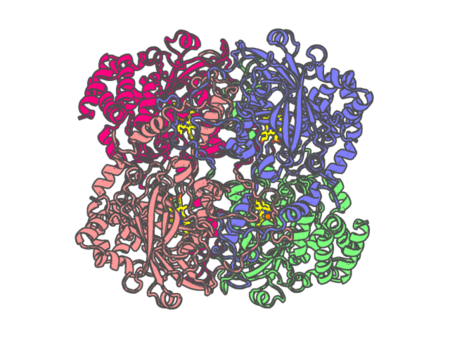 |
Experimental method | X-RAY DIFFRACTION |
|---|---|---|
| Structural data | available | |
| Non-standard amino acids | None | |
| Missing residues/ Structural gap | None / None | |
| Resolution | 2.98 Å | |
| Deposition date | 2004-06-01 |
| pdb_00001thb | REFINEMENT OF A PARTIALLY OXYGENATED T STATE HAEMOGLOBIN AT 1.5 ANGSTROMS RESOLUTION | ||
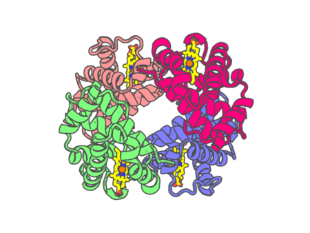 |
Experimental method | X-RAY DIFFRACTION |
|---|---|---|
| Structural data | available | |
| Non-standard amino acids | None | |
| Missing residues/ Structural gap | None / None | |
| Resolution | 1.5 Å | |
| Deposition date | 1990-01-23 |
| pdb_00001tkw | The transient complex of poplar plastocyanin with turnip cytochrome f determined with paramagnetic NMR | ||
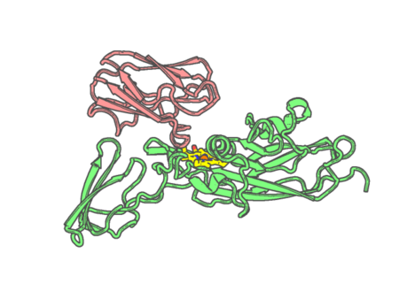 |
Experimental method | SOLUTION NMR |
|---|---|---|
| Structural data | available | |
| Non-standard amino acids | None | |
| Missing residues/ Structural gap | None / None | |
| Resolution | None | |
| Deposition date | 2004-06-09 |
| pdb_00001tqn | Crystal Structure of Human Microsomal P450 3A4 | ||
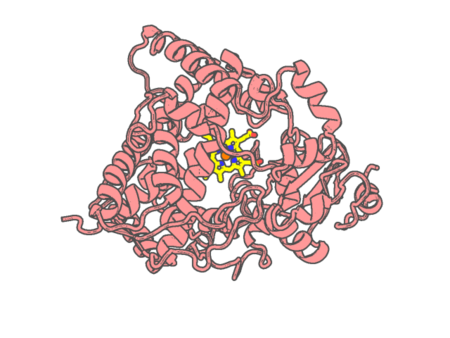 |
Experimental method | X-RAY DIFFRACTION |
|---|---|---|
| Structural data | available | |
| Non-standard amino acids | None | |
| Missing residues/ Structural gap | Exists / Exists | |
| Resolution | 2.05 Å | |
| Deposition date | 2004-06-17 |
| pdb_00001tu2 | THE COMPLEX OF NOSTOC CYTOCHROME F AND PLASTOCYANIN DETERMIN WITH PARAMAGNETIC NMR. BASED ON THE STRUCTURES OF CYTOCHROME F AND PLASTOCYANIN, 10 STRUCTURES | ||
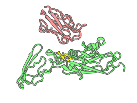 |
Experimental method | SOLUTION NMR |
|---|---|---|
| Structural data | available | |
| Non-standard amino acids | None | |
| Missing residues/ Structural gap | None / None | |
| Resolution | None | |
| Deposition date | 2004-06-24 |
| pdb_00001tu9 | Crystal Structure of a Protein PA3967, a Structurally Highly Homologous to a Human Hemoglobin, from Pseudomonas aeruginosa PAO1 | ||
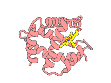 |
Experimental method | X-RAY DIFFRACTION |
|---|---|---|
| Structural data | available | |
| Non-standard amino acids | None | |
| Missing residues/ Structural gap | None / None | |
| Resolution | 1.2 Å | |
| Deposition date | 2004-06-24 |
| pdb_00001u0x | Crystal structure of nitrophorin 4 under pressure of xenon (200 psi) | ||
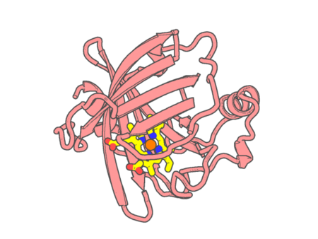 |
Experimental method | X-RAY DIFFRACTION |
|---|---|---|
| Structural data | available | |
| Non-standard amino acids | None | |
| Missing residues/ Structural gap | None / None | |
| Resolution | 1.45 Å | |
| Deposition date | 2004-07-14 |