
Number of data: 3898
Dataset: (exptl_method: X-ray) and (resolution_limit=2.0)
| pdb_00002zwt | Crystal Structure of Ferric Cytochrome P450cam | ||
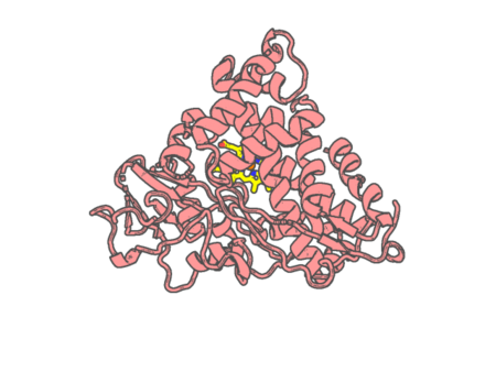 |
Experimental method | X-RAY DIFFRACTION |
|---|---|---|
| Structural data | available | |
| Non-standard amino acids | None | |
| Missing residues/ Structural gap | None / None | |
| Resolution | 1.35 Å | |
| Deposition date | 2008-12-18 |
| pdb_00002zwu | Crystal Structure of Camphor Soaked Ferric Cytochrome P450cam | ||
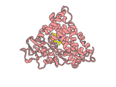 |
Experimental method | X-RAY DIFFRACTION |
|---|---|---|
| Structural data | available | |
| Non-standard amino acids | None | |
| Missing residues/ Structural gap | None / None | |
| Resolution | 1.3 Å | |
| Deposition date | 2008-12-18 |
| pdb_00002zxy | Crystal Structure of Cytochrome c555 from Aquifex aeolicus | ||
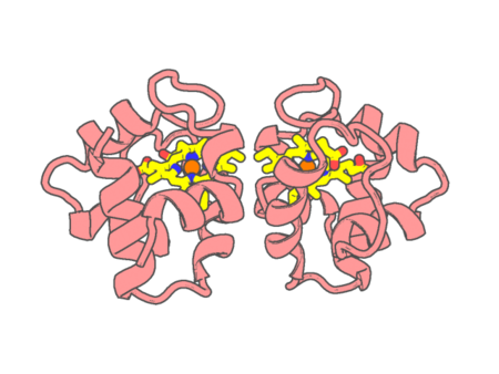 |
Experimental method | X-RAY DIFFRACTION |
|---|---|---|
| Structural data | available | |
| Non-standard amino acids | None | |
| Missing residues/ Structural gap | None / None | |
| Resolution | 1.15 Å | |
| Deposition date | 2009-01-09 |
| pdb_00002zzs | Crystal structure of cytochrome c554 from Vibrio parahaemolyticus strain RIMD2210633 | ||
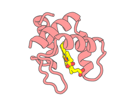 |
Experimental method | X-RAY DIFFRACTION |
|---|---|---|
| Structural data | available | |
| Non-standard amino acids | None | |
| Missing residues/ Structural gap | None / None | |
| Resolution | 1.8 Å | |
| Deposition date | 2009-02-24 |
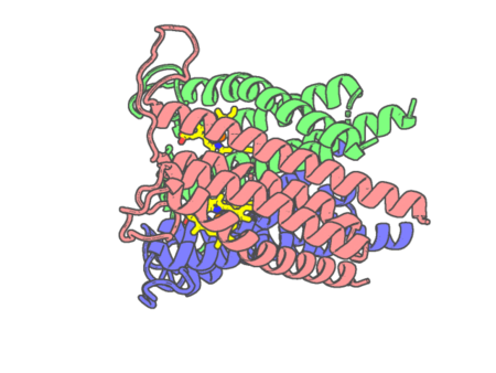
| pdb_000030tw | Crystal structure of Sphingobium lactosutens styrene-oxide isomerase with unknown ligand | ||
 |
Experimental method | X-RAY DIFFRACTION |
|---|---|---|
| Structural data | available | |
| Non-standard amino acids | None | |
| Missing residues/ Structural gap | None / None | |
| Resolution | 1.696 Å | |
| Deposition date | 2026-05-13 |
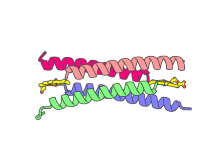
| pdb_000032mq | Crystal structure of the de novo heme-binding peptide GRW-L16CL30H | ||
 |
Experimental method | X-RAY DIFFRACTION |
|---|---|---|
| Structural data | available | |
| Non-standard amino acids | None | |
| Missing residues/ Structural gap | None / None | |
| Resolution | 1.9 Å | |
| Deposition date | 2026-07-16 |
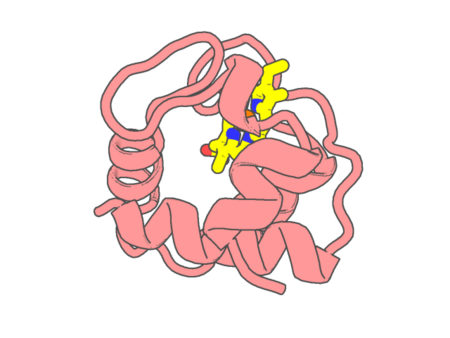
| pdb_0000351c | STRUCTURE OF CYTOCHROME C551 FROM P. AERUGINOSA REFINED AT 1.6 ANGSTROMS RESOLUTION AND COMPARISON OF THE TWO REDOX FORMS | ||
 |
Experimental method | X-RAY DIFFRACTION |
|---|---|---|
| Structural data | available | |
| Non-standard amino acids | None | |
| Missing residues/ Structural gap | None / None | |
| Resolution | 1.6 Å | |
| Deposition date | 1981-07-20 |
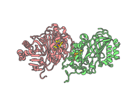
| pdb_00003a15 | Crystal Structure of Substrate-Free Form of Aldoxime Dehydratase (OxdRE) | ||
 |
Experimental method | X-RAY DIFFRACTION |
|---|---|---|
| Structural data | available | |
| Non-standard amino acids | None | |
| Missing residues/ Structural gap | None / None | |
| Resolution | 1.79 Å | |
| Deposition date | 2009-03-26 |
| pdb_00003a16 | Crystal Structure of Aldoxime Dehydratase (OxdRE) in Complex with Propionaldoxime | ||
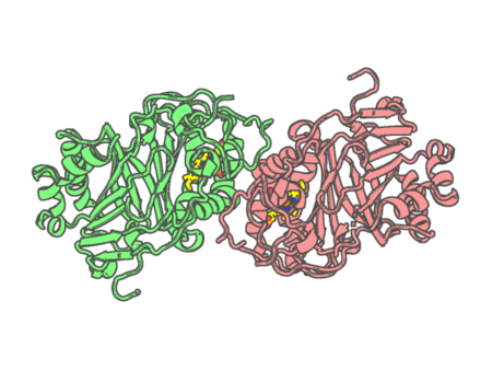 |
Experimental method | X-RAY DIFFRACTION |
|---|---|---|
| Structural data | available | |
| Non-standard amino acids | None | |
| Missing residues/ Structural gap | None / None | |
| Resolution | 1.6 Å | |
| Deposition date | 2009-03-26 |
| pdb_00003a18 | Crystal Structure of Aldoxime Dehydratase (OxdRE) in Complex with Butyraldoxime (soaked crystal) | ||
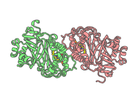 |
Experimental method | X-RAY DIFFRACTION |
|---|---|---|
| Structural data | available | |
| Non-standard amino acids | None | |
| Missing residues/ Structural gap | None / None | |
| Resolution | 1.8 Å | |
| Deposition date | 2009-03-26 |
| pdb_00003a2g | Crystal Structure of K102C-Myoglobin conjugated with Fluorescein | ||
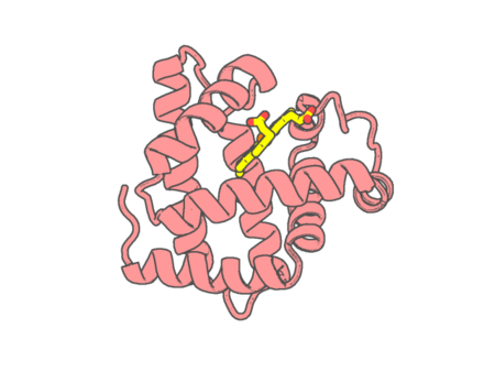 |
Experimental method | X-RAY DIFFRACTION |
|---|---|---|
| Structural data | available | |
| Non-standard amino acids | None | |
| Missing residues/ Structural gap | None / None | |
| Resolution | 1.75 Å | |
| Deposition date | 2009-05-20 |
| pdb_00003a4g | Structure of cytochrome P450 vdh from Pseudonocardia autotrophica (trigonal crystal form) | ||
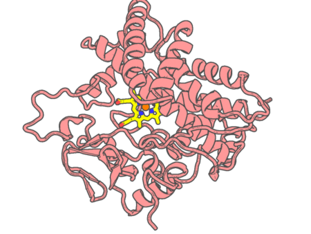 |
Experimental method | X-RAY DIFFRACTION |
|---|---|---|
| Structural data | available | |
| Non-standard amino acids | None | |
| Missing residues/ Structural gap | None / None | |
| Resolution | 1.75 Å | |
| Deposition date | 2009-07-07 |
| pdb_00003a51 | Structure of cytochrome P450 Vdh mutant (Vdh-K1) obtained by directed evolution with bound 25-hydroxyvitamin D3 | ||
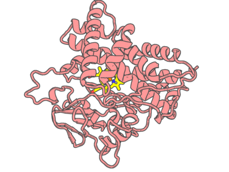 |
Experimental method | X-RAY DIFFRACTION |
|---|---|---|
| Structural data | available | |
| Non-standard amino acids | None | |
| Missing residues/ Structural gap | None / None | |
| Resolution | 2 Å | |
| Deposition date | 2009-07-24 |
| pdb_00003a5a | Crystal structure of a hemoglobin component V from Propsilocerus akamusi (pH5.6 coordinates) | ||
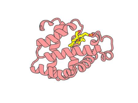 |
Experimental method | X-RAY DIFFRACTION |
|---|---|---|
| Structural data | available | |
| Non-standard amino acids | None | |
| Missing residues/ Structural gap | None / None | |
| Resolution | 1.83 Å | |
| Deposition date | 2009-08-05 |
| pdb_00003a5b | Crystal structure of a hemoglobin component V from Propsilocerus akamusi (pH6.5 coordinates) | ||
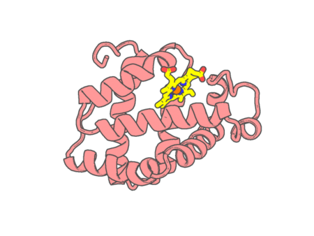 |
Experimental method | X-RAY DIFFRACTION |
|---|---|---|
| Structural data | available | |
| Non-standard amino acids | None | |
| Missing residues/ Structural gap | None / None | |
| Resolution | 1.81 Å | |
| Deposition date | 2009-08-05 |
| pdb_00003a5g | Crystal structure of a hemoglobin component V from Propsilocerus akamusi (pH7.0 coordinates) | ||
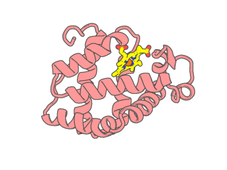 |
Experimental method | X-RAY DIFFRACTION |
|---|---|---|
| Structural data | available | |
| Non-standard amino acids | None | |
| Missing residues/ Structural gap | None / None | |
| Resolution | 1.81 Å | |
| Deposition date | 2009-08-06 |
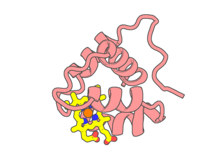
| pdb_00003a9f | Crystal structure of the C-terminal domain of cytochrome cz from Chlorobium tepidum | ||
 |
Experimental method | X-RAY DIFFRACTION |
|---|---|---|
| Structural data | available | |
| Non-standard amino acids | None | |
| Missing residues/ Structural gap | None / None | |
| Resolution | 1.3 Å | |
| Deposition date | 2009-10-25 |
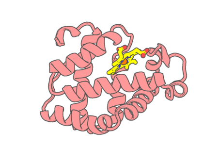
| pdb_00003a9m | Crystal structure of a hemoglobin component V from Propsilocerus akamusi (pH9.0 coordinates) | ||
 |
Experimental method | X-RAY DIFFRACTION |
|---|---|---|
| Structural data | available | |
| Non-standard amino acids | None | |
| Missing residues/ Structural gap | None / None | |
| Resolution | 1.8 Å | |
| Deposition date | 2009-10-30 |
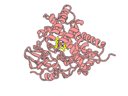
| pdb_00003aba | Crystal structure of CYP105P1 in complex with filipin I | ||
 |
Experimental method | X-RAY DIFFRACTION |
|---|---|---|
| Structural data | available | |
| Non-standard amino acids | None | |
| Missing residues/ Structural gap | None / None | |
| Resolution | 1.8 Å | |
| Deposition date | 2009-12-04 |
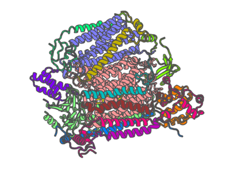
| pdb_00003abk | Bovine heart cytochrome c oxidase at the NO-bound fully reduced state (50K) | ||
 |
Experimental method | X-RAY DIFFRACTION |
|---|---|---|
| Structural data | available | |
| Non-standard amino acids | Included | |
| Missing residues/ Structural gap | None / None | |
| Resolution | 2 Å | |
| Deposition date | 2009-12-16 |