
Number of data: 7660
Dataset: all data
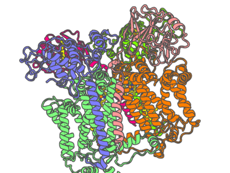
| pdb_000029ya | Cryo-EM structure of Rhodobacter capsulatus cytochrome bc1 dimer with one Rieske protein in the c position and one in the b position and quinone in the quinone oxidation site | ||
 |
Experimental method | ELECTRON MICROSCOPY |
|---|---|---|
| Structural data | available | |
| Non-standard amino acids | None | |
| Missing residues/ Structural gap | Exists / Exists | |
| Resolution | 2.69 Å | |
| Deposition date | 2026-04-14 |
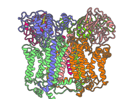
| pdb_000029yb | Cryo-EM structure of Rhodobacter capsulatus cytochrome bc1 dimer with both Rieske proteins in the b position | ||
 |
Experimental method | ELECTRON MICROSCOPY |
|---|---|---|
| Structural data | available | |
| Non-standard amino acids | None | |
| Missing residues/ Structural gap | Exists / Exists | |
| Resolution | None | |
| Deposition date | 2026-04-14 |
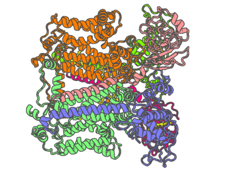
| pdb_000029yc | Cryo-EM structure of Rhodobacter capsulatus cytochrome bc1 dimer with both Rieske proteins in the c position | ||
 |
Experimental method | ELECTRON MICROSCOPY |
|---|---|---|
| Structural data | available | |
| Non-standard amino acids | None | |
| Missing residues/ Structural gap | Exists / Exists | |
| Resolution | 2.43 Å | |
| Deposition date | 2026-04-14 |
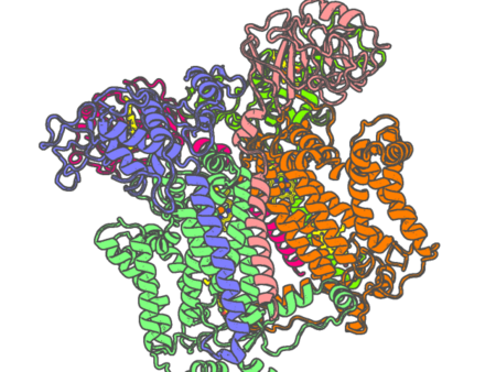
| pdb_000029yd | Cryo-EM structure of Rhodobacter capsulatus cytochrome bc1 dimer with one Rieske protein in the c position and one in an intermediate position | ||
 |
Experimental method | ELECTRON MICROSCOPY |
|---|---|---|
| Structural data | available | |
| Non-standard amino acids | None | |
| Missing residues/ Structural gap | Exists / Exists | |
| Resolution | None | |
| Deposition date | 2026-04-14 |
| pdb_000029ye | Cryo-EM structure of +2Ala insertion mutant of Rhodobacter capsulatus cytochrome bc1 dimer at 2.41 A with quinone in quinone oxidation and reduction sites | ||
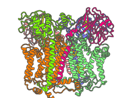 |
Experimental method | ELECTRON MICROSCOPY |
|---|---|---|
| Structural data | available | |
| Non-standard amino acids | None | |
| Missing residues/ Structural gap | Exists / Exists | |
| Resolution | 2.41 Å | |
| Deposition date | 2026-04-14 |
| pdb_00002a06 | Bovine cytochrome bc1 complex with stigmatellin bound | ||
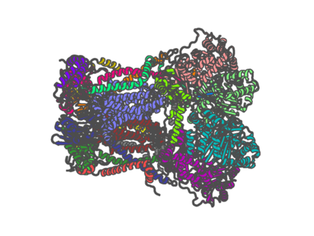 |
Experimental method | X-RAY DIFFRACTION |
|---|---|---|
| Structural data | available | |
| Non-standard amino acids | None | |
| Missing residues/ Structural gap | Exists / Exists | |
| Resolution | 2.1 Å | |
| Deposition date | 2005-06-16 |
| pdb_00002a1m | Crystal structure of ferrous dioxygen complex of wild-type cytochrome P450cam | ||
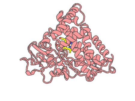 |
Experimental method | X-RAY DIFFRACTION |
|---|---|---|
| Structural data | available | |
| Non-standard amino acids | None | |
| Missing residues/ Structural gap | None / None | |
| Resolution | 2.1 Å | |
| Deposition date | 2005-06-20 |
| pdb_00002a1n | Crystal structure of ferrous dioxygen complex of D251N cytochrome P450cam | ||
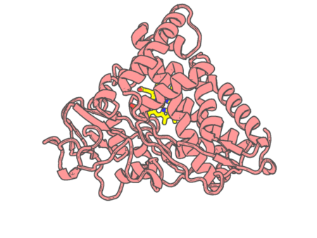 |
Experimental method | X-RAY DIFFRACTION |
|---|---|---|
| Structural data | available | |
| Non-standard amino acids | None | |
| Missing residues/ Structural gap | None / None | |
| Resolution | 1.9 Å | |
| Deposition date | 2005-06-20 |
| pdb_00002a1o | Crystal structure of ferrous dioxygen complex of T252A cytochrome P450cam | ||
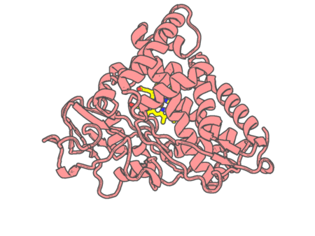 |
Experimental method | X-RAY DIFFRACTION |
|---|---|---|
| Structural data | available | |
| Non-standard amino acids | None | |
| Missing residues/ Structural gap | None / None | |
| Resolution | 1.55 Å | |
| Deposition date | 2005-06-20 |
| pdb_00002a3f | Crystal structure of nitrophorin 2 aqua complex | ||
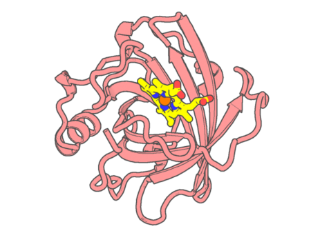 |
Experimental method | X-RAY DIFFRACTION |
|---|---|---|
| Structural data | available | |
| Non-standard amino acids | None | |
| Missing residues/ Structural gap | None / None | |
| Resolution | 1.4 Å | |
| Deposition date | 2005-06-24 |
| pdb_00002a3m | Structure of Desulfovibrio desulfuricans G20 tetraheme cytochrome (oxidized form) | ||
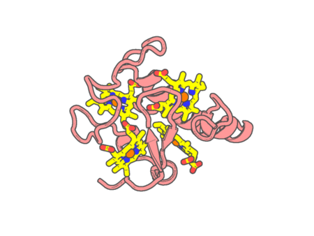 |
Experimental method | X-RAY DIFFRACTION |
|---|---|---|
| Structural data | available | |
| Non-standard amino acids | None | |
| Missing residues/ Structural gap | None / None | |
| Resolution | 1.5 Å | |
| Deposition date | 2005-06-25 |
| pdb_00002a3p | Structure of Desulfovibrio desulfuricans G20 tetraheme cytochrome with bound molybdate | ||
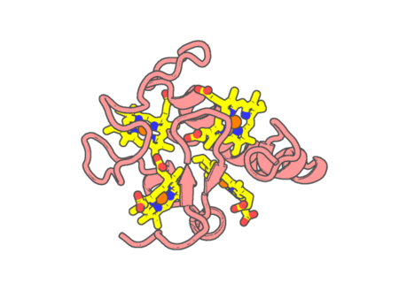 |
Experimental method | X-RAY DIFFRACTION |
|---|---|---|
| Structural data | available | |
| Non-standard amino acids | None | |
| Missing residues/ Structural gap | None / None | |
| Resolution | 2.3 Å | |
| Deposition date | 2005-06-25 |
| pdb_00002a9e | Helicobacter pylori catalase compound I | ||
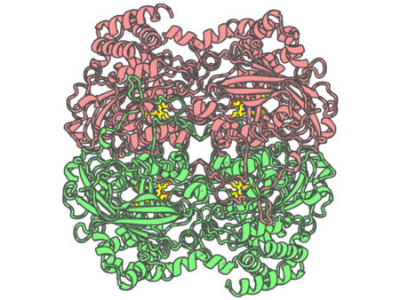 |
Experimental method | X-RAY DIFFRACTION |
|---|---|---|
| Structural data | available | |
| Non-standard amino acids | Included | |
| Missing residues/ Structural gap | None / None | |
| Resolution | 1.76 Å | |
| Deposition date | 2005-07-11 |
| pdb_00002aa1 | Crystal structure of the cathodic hemoglobin isolated from the Antarctic fish Trematomus Newnesi | ||
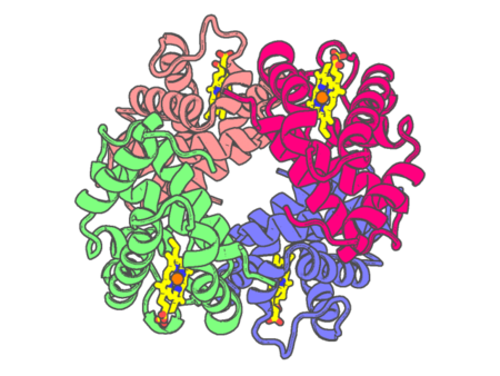 |
Experimental method | X-RAY DIFFRACTION |
|---|---|---|
| Structural data | available | |
| Non-standard amino acids | None | |
| Missing residues/ Structural gap | None / None | |
| Resolution | 1.8 Å | |
| Deposition date | 2005-07-13 |
| pdb_00002acp | Crystal structure of nitrophorin 2 aqua complex | ||
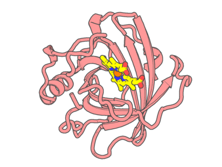 |
Experimental method | X-RAY DIFFRACTION |
|---|---|---|
| Structural data | available | |
| Non-standard amino acids | None | |
| Missing residues/ Structural gap | None / None | |
| Resolution | 1.4 Å | |
| Deposition date | 2005-07-19 |
| pdb_00002acz | Complex II (Succinate Dehydrogenase) From E. Coli with Atpenin A5 inhibitor co-crystallized at the ubiquinone binding site | ||
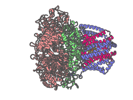 |
Experimental method | X-RAY DIFFRACTION |
|---|---|---|
| Structural data | available | |
| Non-standard amino acids | None | |
| Missing residues/ Structural gap | None / None | |
| Resolution | 3.1 Å | |
| Deposition date | 2005-07-19 |
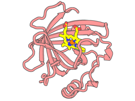
| pdb_00002ah7 | Crystal structure of nitrophorin 2 aqua complex | ||
 |
Experimental method | X-RAY DIFFRACTION |
|---|---|---|
| Structural data | available | |
| Non-standard amino acids | None | |
| Missing residues/ Structural gap | None / None | |
| Resolution | 1.7 Å | |
| Deposition date | 2005-07-27 |
| pdb_00002ai5 | Solution Structure of Cytochrome C552, determined by Distributed Computing Implementation for NMR data | ||
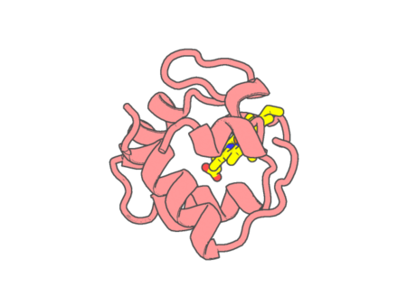 |
Experimental method | SOLUTION NMR |
|---|---|---|
| Structural data | available | |
| Non-standard amino acids | None | |
| Missing residues/ Structural gap | None / None | |
| Resolution | None | |
| Deposition date | 2005-07-29 |
| pdb_00002aiu | Crystal Structure of Mouse Testicular Cytochrome C at 1.6 Angstrom | ||
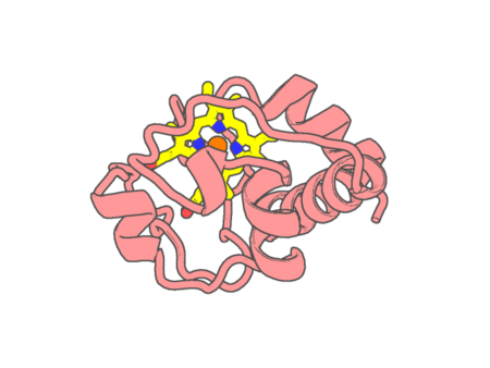 |
Experimental method | X-RAY DIFFRACTION |
|---|---|---|
| Structural data | available | |
| Non-standard amino acids | None | |
| Missing residues/ Structural gap | None / None | |
| Resolution | 1.6 Å | |
| Deposition date | 2005-08-01 |
| pdb_00002al0 | Crystal structure of nitrophorin 2 ferrous aqua complex | ||
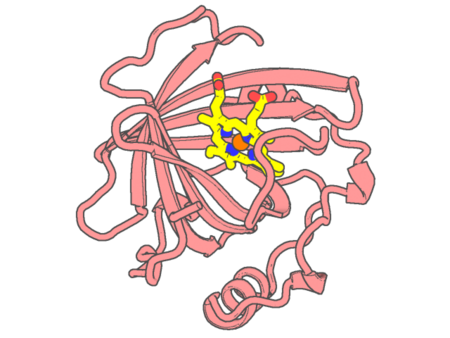 |
Experimental method | X-RAY DIFFRACTION |
|---|---|---|
| Structural data | available | |
| Non-standard amino acids | None | |
| Missing residues/ Structural gap | None / None | |
| Resolution | 1.6 Å | |
| Deposition date | 2005-08-04 |