
Number of data: 7660
Dataset: all data
| pdb_00002pcc | CRYSTAL STRUCTURE OF A COMPLEX BETWEEN ELECTRON TRANSFER PARTNERS, CYTOCHROME C PEROXIDASE AND CYTOCHROME C | ||
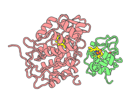 |
Experimental method | X-RAY DIFFRACTION |
|---|---|---|
| Structural data | available | |
| Non-standard amino acids | None | |
| Missing residues/ Structural gap | None / None | |
| Resolution | 2.3 Å | |
| Deposition date | 1993-04-14 |
| pdb_00002pcf | THE COMPLEX OF CYTOCHROME F AND PLASTOCYANIN DETERMINED WITH PARAMAGNETIC NMR. BASED ON THE STRUCTURES OF CYTOCHROME F AND PLASTOCYANIN, 10 STRUCTURES | ||
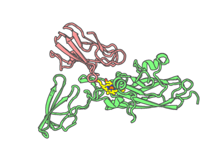 |
Experimental method | SOLUTION NMR |
|---|---|---|
| Structural data | available | |
| Non-standard amino acids | None | |
| Missing residues/ Structural gap | None / None | |
| Resolution | None | |
| Deposition date | 1997-12-22 |
| pdb_00002peg | Crystal structure of Trematomus bernacchii hemoglobin in a partial hemichrome state | ||
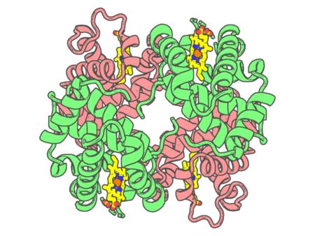 |
Experimental method | X-RAY DIFFRACTION |
|---|---|---|
| Structural data | available | |
| Non-standard amino acids | None | |
| Missing residues/ Structural gap | Exists / Exists | |
| Resolution | 1.48 Å | |
| Deposition date | 2007-04-03 |
| pdb_00002pg5 | Crystal Structure of Human Microsomal P450 2A6 N297Q | ||
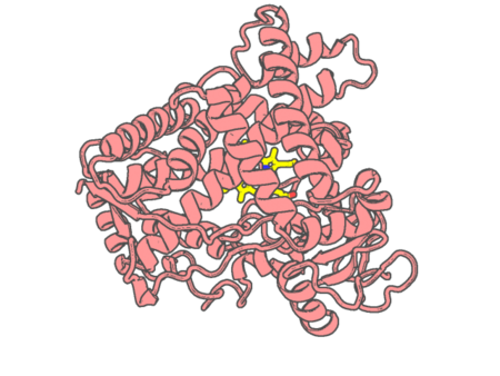 |
Experimental method | X-RAY DIFFRACTION |
|---|---|---|
| Structural data | available | |
| Non-standard amino acids | None | |
| Missing residues/ Structural gap | None / None | |
| Resolution | 1.95 Å | |
| Deposition date | 2007-04-06 |
| pdb_00002pg6 | Crystal Structure of Human Microsomal P450 2A6 L240C/N297Q | ||
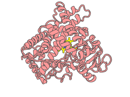 |
Experimental method | X-RAY DIFFRACTION |
|---|---|---|
| Structural data | available | |
| Non-standard amino acids | None | |
| Missing residues/ Structural gap | None / None | |
| Resolution | 2.53 Å | |
| Deposition date | 2007-04-06 |
| pdb_00002pg7 | Crystal Structure of Human Microsomal P450 2A6 N297Q/I300V | ||
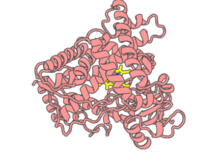 |
Experimental method | X-RAY DIFFRACTION |
|---|---|---|
| Structural data | available | |
| Non-standard amino acids | None | |
| Missing residues/ Structural gap | None / None | |
| Resolution | 2.8 Å | |
| Deposition date | 2007-04-06 |
| pdb_00002pgh | STRUCTURE DETERMINATION OF AQUOMET PORCINE HEMOGLOBIN AT 2.8 ANGSTROM RESOLUTION | ||
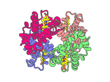 |
Experimental method | X-RAY DIFFRACTION |
|---|---|---|
| Structural data | available | |
| Non-standard amino acids | None | |
| Missing residues/ Structural gap | None / None | |
| Resolution | 2.8 Å | |
| Deposition date | 1994-09-16 |
| pdb_00002prc | PHOTOSYNTHETIC REACTION CENTER FROM RHODOPSEUDOMONAS VIRIDIS (UBIQUINONE-2 COMPLEX) | ||
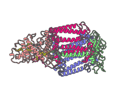 |
Experimental method | X-RAY DIFFRACTION |
|---|---|---|
| Structural data | available | |
| Non-standard amino acids | Included | |
| Missing residues/ Structural gap | None / None | |
| Resolution | 2.45 Å | |
| Deposition date | 1997-07-29 |
| pdb_00002pt3 | Crystal structure of bovine lactoperoxidase at 2.34 A resolution reveals multiple anion binding sites | ||
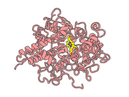 |
Experimental method | X-RAY DIFFRACTION |
|---|---|---|
| Structural data | available | |
| Non-standard amino acids | Included | |
| Missing residues/ Structural gap | None / None | |
| Resolution | 2.34 Å | |
| Deposition date | 2007-05-08 |
| pdb_00002pum | Crystal structure of bovine lactoperoxidase complex with catechol and iodide at 2.7 A resolution | ||
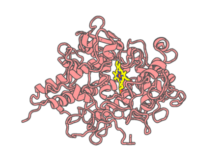 |
Experimental method | X-RAY DIFFRACTION |
|---|---|---|
| Structural data | available | |
| Non-standard amino acids | Included | |
| Missing residues/ Structural gap | None / None | |
| Resolution | 2.7 Å | |
| Deposition date | 2007-05-09 |
| pdb_00002q6n | Structure of Cytochrome P450 2B4 with Bound 1-(4-cholorophenyl)imidazole | ||
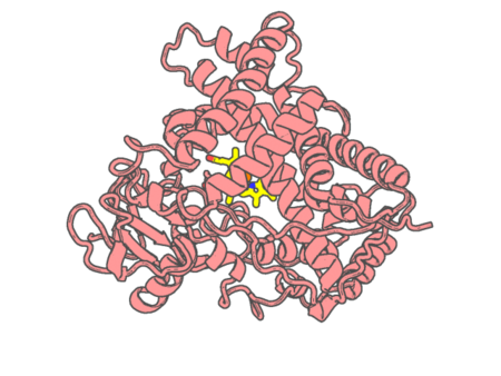 |
Experimental method | X-RAY DIFFRACTION |
|---|---|---|
| Structural data | available | |
| Non-standard amino acids | None | |
| Missing residues/ Structural gap | None / None | |
| Resolution | 3.2 Å | |
| Deposition date | 2007-06-05 |
| pdb_00002q7a | Crystal structure of the cell surface heme transfer protein Shp | ||
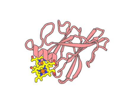 |
Experimental method | X-RAY DIFFRACTION |
|---|---|---|
| Structural data | available | |
| Non-standard amino acids | None | |
| Missing residues/ Structural gap | None / None | |
| Resolution | 2.1 Å | |
| Deposition date | 2007-06-06 |
| pdb_00002q8p | Crystal Structure of selenomethionine labelled S. aureus IsdE complexed with heme | ||
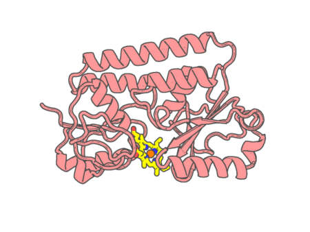 |
Experimental method | X-RAY DIFFRACTION |
|---|---|---|
| Structural data | available | |
| Non-standard amino acids | Included | |
| Missing residues/ Structural gap | None / None | |
| Resolution | 1.95 Å | |
| Deposition date | 2007-06-11 |
| pdb_00002q8q | Crystal Structure of S. aureus IsdE complexed with heme | ||
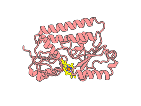 |
Experimental method | X-RAY DIFFRACTION |
|---|---|---|
| Structural data | available | |
| Non-standard amino acids | None | |
| Missing residues/ Structural gap | None / None | |
| Resolution | 2.15 Å | |
| Deposition date | 2007-06-11 |
| pdb_00002q9f | Crystal structure of human cytochrome P450 46A1 in complex with cholesterol-3-sulphate | ||
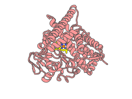 |
Experimental method | X-RAY DIFFRACTION |
|---|---|---|
| Structural data | available | |
| Non-standard amino acids | None | |
| Missing residues/ Structural gap | None / None | |
| Resolution | 1.9 Å | |
| Deposition date | 2007-06-12 |
| pdb_00002q9g | Crystal structure of human cytochrome P450 46A1 | ||
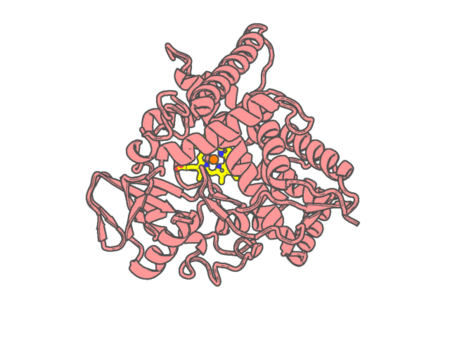 |
Experimental method | X-RAY DIFFRACTION |
|---|---|---|
| Structural data | available | |
| Non-standard amino acids | None | |
| Missing residues/ Structural gap | Exists / Exists | |
| Resolution | 2.4 Å | |
| Deposition date | 2007-06-12 |
| pdb_00002qbl | Crystal structure of ferric G248T cytochrome P450cam | ||
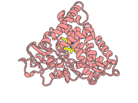 |
Experimental method | X-RAY DIFFRACTION |
|---|---|---|
| Structural data | available | |
| Non-standard amino acids | None | |
| Missing residues/ Structural gap | None / None | |
| Resolution | 1.8 Å | |
| Deposition date | 2007-06-18 |
| pdb_00002qbm | Crystal structure of the P450cam G248T mutant in the cyanide bound state | ||
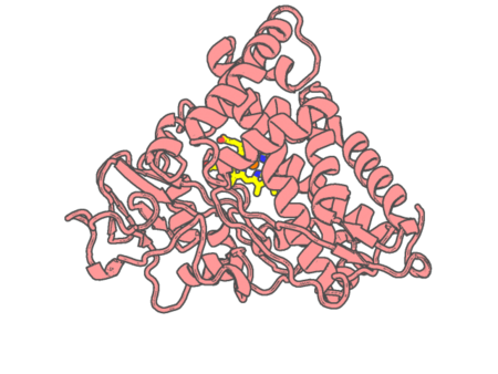 |
Experimental method | X-RAY DIFFRACTION |
|---|---|---|
| Structural data | available | |
| Non-standard amino acids | None | |
| Missing residues/ Structural gap | None / None | |
| Resolution | 1.8 Å | |
| Deposition date | 2007-06-18 |
| pdb_00002qbn | Crystal structure of ferric G248V cytochrome P450cam | ||
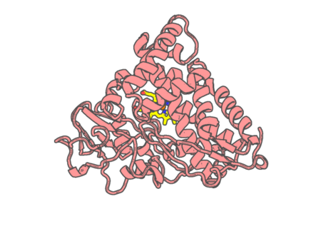 |
Experimental method | X-RAY DIFFRACTION |
|---|---|---|
| Structural data | available | |
| Non-standard amino acids | None | |
| Missing residues/ Structural gap | None / None | |
| Resolution | 1.75 Å | |
| Deposition date | 2007-06-18 |
| pdb_00002qbo | Crystal structure of the P450cam G248V mutant in the cyanide bound state | ||
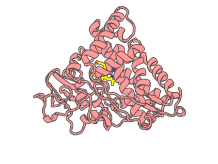 |
Experimental method | X-RAY DIFFRACTION |
|---|---|---|
| Structural data | available | |
| Non-standard amino acids | None | |
| Missing residues/ Structural gap | None / None | |
| Resolution | 1.9 Å | |
| Deposition date | 2007-06-18 |