
Number of data: 3440
Dataset: (without mutations)
| pdb_00005agl | Structure of rat neuronal nitric oxide synthase heme domain in complex with (S)-2-Amino-5-(2-(methylsulfonyl)acetimidamido)pentanoic acid | ||
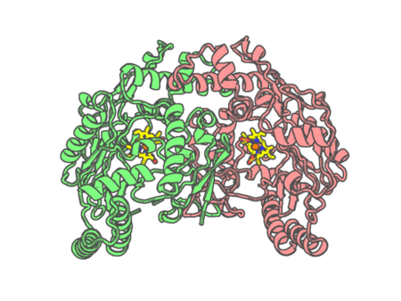 |
Experimental method | X-RAY DIFFRACTION |
|---|---|---|
| Structural data | available | |
| Non-standard amino acids | None | |
| Missing residues/ Structural gap | Exists / Exists | |
| Resolution | 1.94 Å | |
| Deposition date | 2015-02-02 |
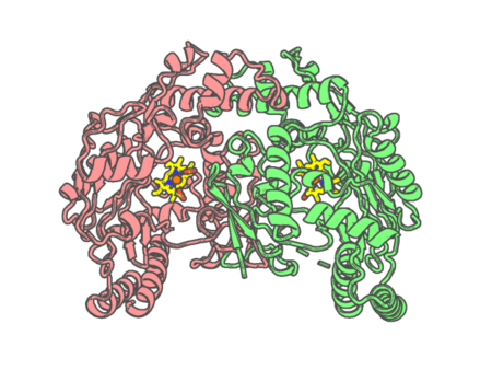
| pdb_00005agm | Structure of rat neuronal nitric oxide synthase heme domain in complex with (S)-2-Amino-5-(2-oxoacetimidamido)pentanoic acid | ||
 |
Experimental method | X-RAY DIFFRACTION |
|---|---|---|
| Structural data | available | |
| Non-standard amino acids | None | |
| Missing residues/ Structural gap | Exists / Exists | |
| Resolution | 1.84 Å | |
| Deposition date | 2015-02-02 |
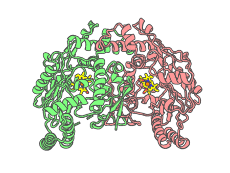
| pdb_00005agn | Structure of rat neuronal nitric oxide synthase heme domain in complex with (S)-2-Amino-5-(2-hydroxyacetimidamido)pentanoic acid | ||
 |
Experimental method | X-RAY DIFFRACTION |
|---|---|---|
| Structural data | available | |
| Non-standard amino acids | None | |
| Missing residues/ Structural gap | Exists / Exists | |
| Resolution | 1.95 Å | |
| Deposition date | 2015-02-02 |
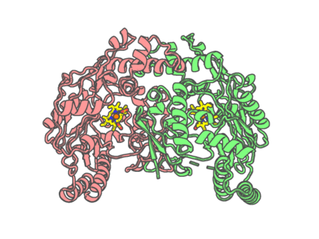
| pdb_00005ago | Structure of rat neuronal nitric oxide synthase heme domain in complex with (S)-2-Amino-5-(2-mercaptoacetimidamido)pentanoic acid | ||
 |
Experimental method | X-RAY DIFFRACTION |
|---|---|---|
| Structural data | available | |
| Non-standard amino acids | None | |
| Missing residues/ Structural gap | Exists / Exists | |
| Resolution | 1.902 Å | |
| Deposition date | 2015-02-02 |
| pdb_00005agp | Structure of rat neuronal nitric oxide synthase heme domain in complex with (S)-2-Amino-5-(2-mercaptoacetimidamido)pentanoic acid | ||
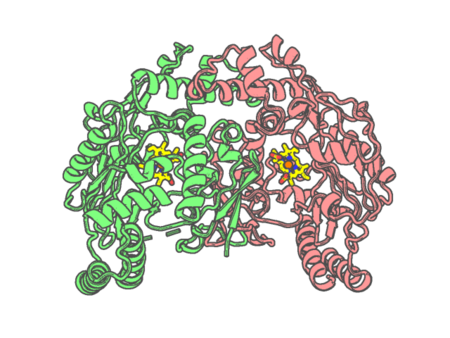 |
Experimental method | X-RAY DIFFRACTION |
|---|---|---|
| Structural data | available | |
| Non-standard amino acids | None | |
| Missing residues/ Structural gap | Exists / Exists | |
| Resolution | 2.1 Å | |
| Deposition date | 2015-02-02 |
| pdb_00005aur | Hydrogenobacter thermophilus cytochrome c552 dimer formed by domain swapping at N-terminal region | ||
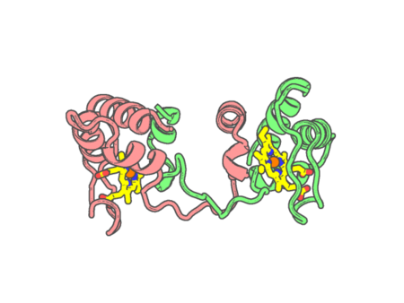 |
Experimental method | X-RAY DIFFRACTION |
|---|---|---|
| Structural data | available | |
| Non-standard amino acids | None | |
| Missing residues/ Structural gap | None / None | |
| Resolution | 1.26 Å | |
| Deposition date | 2015-06-08 |
| pdb_00005aus | Hydrogenobacter thermophilus cytochrome c552 dimer formed by domain swapping at C-terminal region | ||
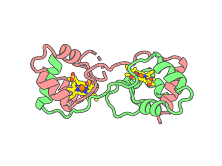 |
Experimental method | X-RAY DIFFRACTION |
|---|---|---|
| Structural data | available | |
| Non-standard amino acids | None | |
| Missing residues/ Structural gap | Exists / Exists | |
| Resolution | 1.3 Å | |
| Deposition date | 2015-06-08 |
| pdb_00005az3 | Crystal structure of heme binding protein HmuT | ||
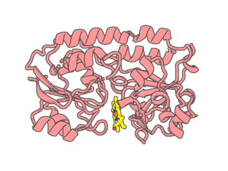 |
Experimental method | X-RAY DIFFRACTION |
|---|---|---|
| Structural data | available | |
| Non-standard amino acids | None | |
| Missing residues/ Structural gap | None / None | |
| Resolution | 1.423 Å | |
| Deposition date | 2015-09-18 |
| pdb_00005b1a | Bovine heart cytochrome c oxidase in the fully oxidized state at 1.5 angstrom resolution | ||
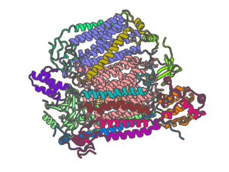 |
Experimental method | X-RAY DIFFRACTION |
|---|---|---|
| Structural data | available | |
| Non-standard amino acids | Included | |
| Missing residues/ Structural gap | None / None | |
| Resolution | 1.5 Å | |
| Deposition date | 2015-12-01 |
| pdb_00005b1b | Bovine heart cytochrome c oxidase in the fully reduced state at 1.6 angstrom resolution | ||
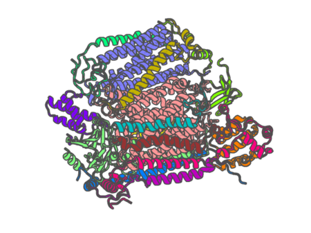 |
Experimental method | X-RAY DIFFRACTION |
|---|---|---|
| Structural data | available | |
| Non-standard amino acids | Included | |
| Missing residues/ Structural gap | None / None | |
| Resolution | 1.6 Å | |
| Deposition date | 2015-12-01 |
| pdb_00005b2v | Crystal Structure of P450BM3 with N-perfluorohexanoyl-L-tryptophan | ||
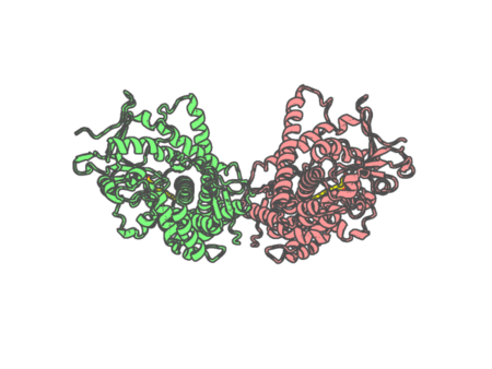 |
Experimental method | X-RAY DIFFRACTION |
|---|---|---|
| Structural data | available | |
| Non-standard amino acids | None | |
| Missing residues/ Structural gap | None / None | |
| Resolution | 2.3 Å | |
| Deposition date | 2016-02-03 |
| pdb_00005b2w | Crystal Structure of P450BM3 with N-perfluorododecanoyl-L-tryptophan | ||
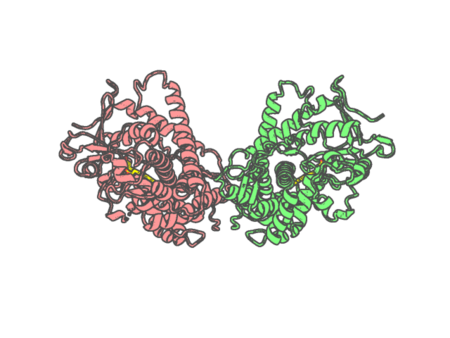 |
Experimental method | X-RAY DIFFRACTION |
|---|---|---|
| Structural data | available | |
| Non-standard amino acids | None | |
| Missing residues/ Structural gap | None / None | |
| Resolution | 1.65 Å | |
| Deposition date | 2016-02-03 |
| pdb_00005b2y | Crystal Structure of P450BM3 with N-perfluorodecanoyl-L-tryptophan | ||
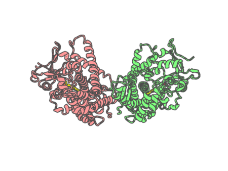 |
Experimental method | X-RAY DIFFRACTION |
|---|---|---|
| Structural data | available | |
| Non-standard amino acids | None | |
| Missing residues/ Structural gap | None / None | |
| Resolution | 2.01 Å | |
| Deposition date | 2016-02-07 |
| pdb_00005b3i | Homo-dimeric structure of cytochrome c' from Thermophilic Hydrogenophilus thermoluteolus | ||
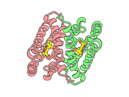 |
Experimental method | X-RAY DIFFRACTION |
|---|---|---|
| Structural data | available | |
| Non-standard amino acids | None | |
| Missing residues/ Structural gap | None / None | |
| Resolution | 1.89 Å | |
| Deposition date | 2016-02-29 |
| pdb_00005b3s | Bovine heart cytochrome c oxidase in the carbon monoxide-bound mixed-valence state at 1.68 angstrom resolution (50 K) | ||
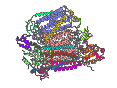 |
Experimental method | X-RAY DIFFRACTION |
|---|---|---|
| Structural data | available | |
| Non-standard amino acids | Included | |
| Missing residues/ Structural gap | None / None | |
| Resolution | 1.68 Å | |
| Deposition date | 2016-03-11 |
| pdb_00005b5m | Crystal structure of the Sr-substituted LH1-RC complex from Tch. tepidum | ||
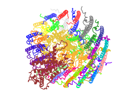 |
Experimental method | X-RAY DIFFRACTION |
|---|---|---|
| Structural data | available | |
| Non-standard amino acids | None | |
| Missing residues/ Structural gap | None / None | |
| Resolution | 3.3 Å | |
| Deposition date | 2016-05-12 |
| pdb_00005b5n | Crystal structure of the Ba-substituted LH1-RC complex from Tch. tepidum | ||
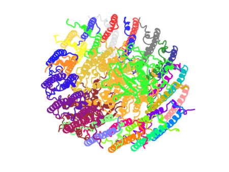 |
Experimental method | X-RAY DIFFRACTION |
|---|---|---|
| Structural data | available | |
| Non-standard amino acids | None | |
| Missing residues/ Structural gap | None / None | |
| Resolution | 3.3 Å | |
| Deposition date | 2016-05-12 |
| pdb_00005b6q | Crystal structure of monomeric cytochrome c5 from Shewanella violacea | ||
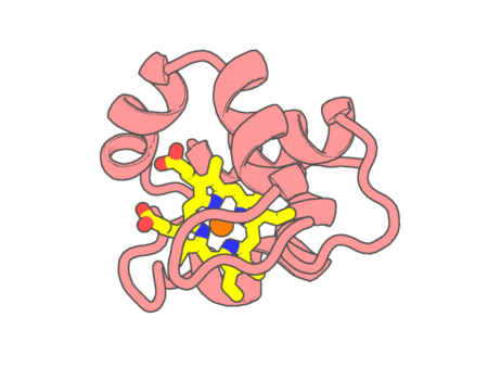 |
Experimental method | X-RAY DIFFRACTION |
|---|---|---|
| Structural data | available | |
| Non-standard amino acids | None | |
| Missing residues/ Structural gap | None / None | |
| Resolution | 1.78 Å | |
| Deposition date | 2016-06-01 |
| pdb_00005b82 | Crystal structure of the cyanobacterial heme-protein Tll0287 | ||
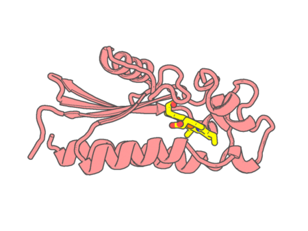 |
Experimental method | X-RAY DIFFRACTION |
|---|---|---|
| Structural data | available | |
| Non-standard amino acids | None | |
| Missing residues/ Structural gap | None / None | |
| Resolution | 2 Å | |
| Deposition date | 2016-06-12 |
| pdb_00005c0z | The structure of oxidized rat cytochrome c at 1.13 angstroms resolution | ||
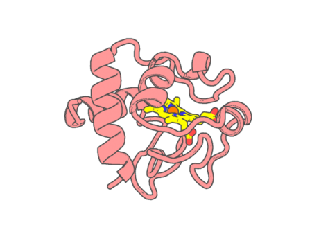 |
Experimental method | X-RAY DIFFRACTION |
|---|---|---|
| Structural data | available | |
| Non-standard amino acids | None | |
| Missing residues/ Structural gap | None / None | |
| Resolution | 1.1236 Å | |
| Deposition date | 2015-06-12 |