PDB ID:  pdb_00001d7d
  • CYTOCHROME DOMAIN OF CELLOBIOSE DEHYDROGENASE, HP3 FRAGMENT, PH 7.5
  • Experimental method: X-RAY DIFFRACTION
  • Resolution: 1.9 Å
  • Deposition date: 1999-10-16
Number of assemblies:  1
Assembly ID:  0  (author_and_software_defined_assembly)    
Structural data modeled
Oligomeric count 2
label_asym_ids of protein
label_asym_ids of heme
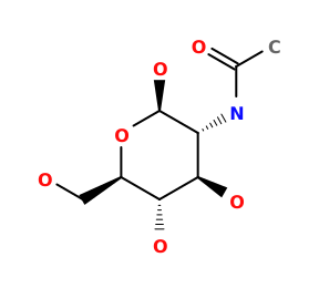
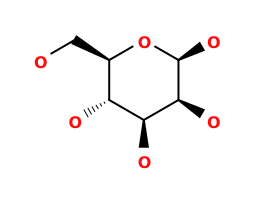
label_asym_ids of the others C (NAGStructure of NAGNAG , BMAStructure of BMABMA ), D (NAGStructure of NAGNAG , MANStructure of MANMAN ), E (CDStructure of CDCD ), F (CDStructure of CDCD ), G (CDStructure of CDCD ), H (CDStructure of CDCD ), J (CDStructure of CDCD ), K (CDStructure of CDCD ), M (1PGStructure of 1PG1PG )
Each chain information
Chain A heme  
auth_asym_id A
Residue indices (Uniprot) 1-186 (19-204)
Missing residues 1GLN-1GLN
Nonstandard amino acids Structure of PCAPCA
Uniprot ID Q01738 →UniprotKB
S-S bonds 121:124
label_asym_ids of heme I
Available structure PDB and Modeled
RMSD of experimental residues after energy minimization 0.09 Å
Chain B heme  
auth_asym_id B
Residue indices (Uniprot) 1-186 (19-204)
Missing residues 1GLN-1GLN
Nonstandard amino acids Structure of PCAPCA
Uniprot ID Q01738 →UniprotKB
S-S bonds 121:124
label_asym_ids of heme L
Available structure PDB and Modeled
RMSD of experimental residues after energy minimization 0.11 Å