PDB ID:  pdb_00001mhl
  • CRYSTAL STRUCTURE OF HUMAN MYELOPEROXIDASE ISOFORM C CRYSTALLIZED IN SPACE GROUP P2(1) AT PH 5.5 AND 20 DEG C
  • Experimental method: X-RAY DIFFRACTION
  • Resolution: 2.25 Å
  • Deposition date: 1995-06-09
Number of assemblies:  1
Assembly ID:  0  (author_and_software_defined_assembly)    
Structural data PDB
Oligomeric count 4
label_asym_ids of protein
label_asym_ids of heme
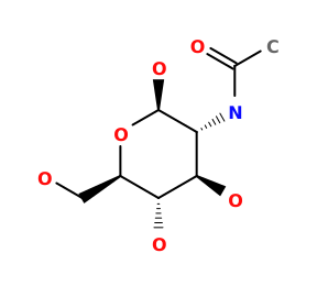
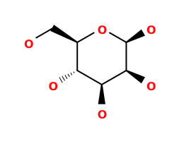
label_asym_ids of the others E (NAGStructure of NAGNAG , BMAStructure of BMABMA , MANStructure of MANMAN , FUCStructure of FUCFUC ), F (NAGStructure of NAGNAG , BMAStructure of BMABMA , MANStructure of MANMAN , FUCStructure of FUCFUC ), G (NAGStructure of NAGNAG ), H (NAGStructure of NAGNAG ), I (CAStructure of CACA ), J (CLStructure of CLCL ), L (NAGStructure of NAGNAG ), M (NAGStructure of NAGNAG ), N (CAStructure of CACA ), O (CLStructure of CLCL )
Each chain information

Showing 2 chains with heme of 4. Show all chains

Chain B heme  
auth_asym_id C
Residue indices (Uniprot) 113-578 (279-744)
Uniprot ID P05164 →UniprotKB
S-S bonds 115:125,119:143,221:232,440:497,538:564
label_asym_ids of heme K
Available structure PDB
Chain D heme  
auth_asym_id D
Residue indices (Uniprot) 113-578 (279-744)
Uniprot ID P05164 →UniprotKB
S-S bonds 115:125,119:143,221:232,440:497,538:564
label_asym_ids of heme P
Available structure PDB