PDB ID: pdb_00002ikc
- Crystal structure of sheep lactoperoxidase at 3.25 A resolution reveals the binding sites for formate
- Experimental method: X-RAY DIFFRACTION
- Resolution: 3.25 Å
- Deposition date: 2006-10-02
Number of assemblies: 1
| Structural data | PDB |
|---|---|
| Oligomeric count | 2 |
| label_asym_ids of protein | |
| label_asym_ids of heme | |
| label_asym_ids of the others |
C (NAG 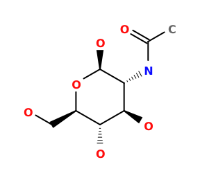 NAG
), D (NAGNAG
), E (NAGNAG
, MAN  MAN
), F (NAGNAG
), G (NAGNAG
), H (NAGNAG
, MANMAN
), I (NAGNAG
), J (CA  CA
), K (CO3  CO3
), M (FMT  FMT
), N (FMTFMT
), O (FMTFMT
), P (NAGNAG
), Q (CACA
), R (CO3CO3
), T (FMTFMT
), U (FMTFMT
), V (FMTFMT
) |

Each chain information
| auth_asym_id | A |
|---|---|
| Residue indices (Uniprot) | 1-595 (118-712) |
| Uniprot ID | A0ABF7PFE0 →UniprotKB |
| S-S bonds | 6:167,15:28,129:139,133:157,237:248,456:513,554:579 |
| label_asym_ids of heme | L |
| Available structure | PDB |
| auth_asym_id | B |
|---|---|
| Residue indices (Uniprot) | 1-595 (118-712) |
| Uniprot ID | A0ABF7PFE0 →UniprotKB |
| S-S bonds | 6:167,15:28,129:139,133:157,237:248,456:513,554:579 |
| label_asym_ids of heme | S |
| Available structure | PDB |