PDB ID: pdb_00004wjg
- Structure of T. brucei haptoglobin-hemoglobin receptor binding to human haptoglobin-hemoglobin
- Experimental method: X-RAY DIFFRACTION
- Resolution: 3.1 Å
- Deposition date: 2014-09-30
Number of assemblies: 3
| Structural data | modeled |
|---|---|
| Oligomeric count | 10 |
| label_asym_ids of protein | |
| label_asym_ids of heme | |
| label_asym_ids of the others |
AB (NAG 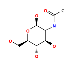 NAG
), BB (NAGNAG
), CB (NAGNAG
), DB (NAGNAG
), EA (NAGNAG
), FA (NAGNAG
), GA (NAGNAG
), PA (OXY  OXY
), RA (OXYOXY
), SA (NAGNAG
), TA (NAGNAG
), UA (NAGNAG
), VA (NAGNAG
), XA (OXYOXY
), ZA (OXYOXY
) |
Each chain information
Showing 4 chains with heme of 10. Show all chains
| auth_asym_id | A |
|---|---|
| Residue indices (Uniprot) | 1-141 (2-142) |
| Uniprot ID | P69905 →UniprotKB |
| label_asym_ids of heme | OA |
| Available structure | PDB |
| auth_asym_id | B |
|---|---|
| Residue indices (Uniprot) | 1-146 (2-147) |
| Uniprot ID | P68871 →UniprotKB |
| label_asym_ids of heme | QA |
| Available structure | PDB |
| Structural data | modeled |
|---|---|
| Oligomeric count | 10 |
| label_asym_ids of protein | |
| label_asym_ids of heme | |
| label_asym_ids of the others |
FB (OXYOXY
), HA (NAGNAG
), HB (OXYOXY
), IA (NAGNAG
), IB (NAGNAG
), JA (NAGNAG
), JB (NAGNAG
), KA (NAGNAG
), KB (NAGNAG
), LB (NAGNAG
), NB (OXYOXY
), PB (OXYOXY
), QB (NAGNAG
), RB (NAGNAG
), SB (NAGNAG
), TB (NAGNAG
)
|
Each chain information
Showing 4 chains with heme of 10. Show all chains
| auth_asym_id | K |
|---|---|
| Residue indices (Uniprot) | 1-141 (2-142) |
| Uniprot ID | P69905 →UniprotKB |
| label_asym_ids of heme | EB |
| Available structure | PDB |
| auth_asym_id | L |
|---|---|
| Residue indices (Uniprot) | 1-146 (2-147) |
| Uniprot ID | P68871 →UniprotKB |
| label_asym_ids of heme | GB |
| Available structure | PDB |
| Structural data | modeled |
|---|---|
| Oligomeric count | 10 |
| label_asym_ids of protein | |
| label_asym_ids of heme | |
| label_asym_ids of the others |
AC (NAGNAG
), BC (NAGNAG
), DC (OXYOXY
), FC (OXYOXY
), GC (NAGNAG
), HC (NAGNAG
), IC (NAGNAG
), LA (NAGNAG
), MA (NAGNAG
), NA (NAGNAG
), VB (OXYOXY
), XB (OXYOXY
), YB (NAGNAG
), ZB (NAGNAG
)
|
Each chain information
Showing 4 chains with heme of 10. Show all chains
| auth_asym_id | 1 |
|---|---|
| Residue indices (Uniprot) | 1-146 (2-147) |
| Uniprot ID | P68871 →UniprotKB |
| label_asym_ids of heme | EC |
| Available structure | PDB |
| auth_asym_id | U |
|---|---|
| Residue indices (Uniprot) | 1-141 (2-142) |
| Uniprot ID | P69905 →UniprotKB |
| label_asym_ids of heme | UB |
| Available structure | PDB |