PDB ID: pdb_00005wv3
- Crystal structure of bovine lactoperoxidase with a partial Glu258-heme linkage at 2.07 A resolution.
- Experimental method: X-RAY DIFFRACTION
- Resolution: 2.07 Å
- Deposition date: 2016-12-21
Number of assemblies: 1
| Structural data | modeled |
|---|---|
| Oligomeric count | 1 |
| label_asym_ids of protein | |
| label_asym_ids of heme | |
| label_asym_ids of the others |
B (NAG 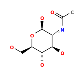 NAG
), C (CA  CA
), E (NAGNAG
), F (NAGNAG
), G (NAGNAG
), H (IOD  IOD
), I (OSM  OSM
), J (OSMOSM
), K (OSMOSM
), L (OSMOSM
), M (OSMOSM
), N (OSMOSM
), O (OSMOSM
), P (OSMOSM
), Q (OSMOSM
), R (GOL  GOL
), S (IODIOD
), T (BR  BR
), U (IODIOD
), V (OSMOSM
), W (GOLGOL
) |

Each chain information
| auth_asym_id | A |
|---|---|
| Residue indices (Uniprot) | 1-595 (118-712) |
| Mutations (UniProt numbering) | |
| Missing residues | 198SER-198SER |
| Nonstandard amino acids |
 SEP |
| Uniprot ID | P80025 →UniprotKB |
| S-S bonds | 6:167,15:28,129:139,133:157,237:248,456:513,554:579 |
| label_asym_ids of heme | D |
| Available structure | PDB and Modeled |
| RMSD of experimental residues after energy minimization | 0.12 Å |