PDB ID: pdb_00009q4k
- Structure of human neuronal nitric oxide synthase R354A/G357D mutant heme domain bound with 6-((2,3-difluoro-5-(2-(piperidin-1-yl)ethyl)phenoxy)methyl)-4-methylpyridin-2-amine
- Experimental method: X-RAY DIFFRACTION
- Resolution: 2.198 Å
- Deposition date: 2025-08-20
Number of assemblies: 2
| Structural data | modeled |
|---|---|
| Oligomeric count | 2 |
| label_asym_ids of protein | |
| label_asym_ids of heme | |
| label_asym_ids of the others |
F (A1C 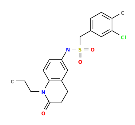 A1C
), G (GOL  GOL
), H (GOLGOL
), I (ZN  ZN
), J (H4B  H4B
), L (H4BH4B
), M (A1CA1C
), N (GOLGOL
) |

Each chain information
| auth_asym_id | A |
|---|---|
| Residue indices (Uniprot) | 304-722 (302-722) |
| Mutations (UniProt numbering) | |
| Missing residues | 344SER-348ARG |
| Structural gaps | Exists |
| Uniprot ID | P29475 →UniprotKB |
| label_asym_ids of heme | E |
| Available structure | PDB and Modeled |
| RMSD of experimental residues after energy minimization | 0.10 Å |
| Structural data | modeled |
|---|---|
| Oligomeric count | 2 |
| label_asym_ids of protein | |
| label_asym_ids of heme | |
| label_asym_ids of the others |
P (H4BH4B
), Q (A1CA1C
), R (GOLGOL
), S (GOLGOL
), T (ZNZN
), V (H4BH4B
), W (A1CA1C
), X (GOLGOL
)
|
Each chain information
| auth_asym_id | C |
|---|---|
| Residue indices (Uniprot) | 304-722 (302-722) |
| Mutations (UniProt numbering) | |
| Uniprot ID | P29475 →UniprotKB |
| label_asym_ids of heme | O |
| Available structure | PDB |
| auth_asym_id | D |
|---|---|
| Residue indices (Uniprot) | 304-722 (302-722) |
| Mutations (UniProt numbering) | |
| Missing residues | 345GLN-351GLU |
| Structural gaps | Exists |
| Uniprot ID | P29475 →UniprotKB |
| label_asym_ids of heme | U |
| Available structure | PDB and Modeled |
| RMSD of experimental residues after energy minimization | 0.12 Å |