PDB ID: pdb_00009qj3
- Structure of native leukocyte myeloperoxidase in complex with a truncated version of the Staphylococcal Peroxidase Inhibitor SPIN and iodide at pH 5.5
- Experimental method: X-RAY DIFFRACTION
- Resolution: 2.63 Å
- Deposition date: 2025-03-18
Number of assemblies: 1
| Structural data | modeled |
|---|---|
| Oligomeric count | 6 |
| label_asym_ids of protein | |
| label_asym_ids of heme | |
| label_asym_ids of the others |
AA (IOD  IOD
), AB (IODIOD
), AC (IODIOD
), AD (IODIOD
), BA (IODIOD
), BB (CA  CA
), BC (IODIOD
), CA (IODIOD
), CC (IODIOD
), DA (IODIOD
), DB (IODIOD
), DC (IODIOD
), EA (IODIOD
), EB (IODIOD
), EC (IODIOD
), FA (IODIOD
), FB (IODIOD
), FC (IODIOD
), G (NAG 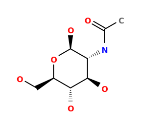 NAG
), GA (IODIOD
), GB (IODIOD
), GC (IODIOD
), H (NAGNAG
), HA (IODIOD
), HB (IODIOD
), HC (IODIOD
), I (NAGNAG
, MAN  MAN
, FUC  FUC
), IA (IODIOD
), IB (IODIOD
), IC (IODIOD
), J (NAGNAG
), JA (IODIOD
), JB (IODIOD
), JC (IODIOD
), K (NAGNAG
), KA (IODIOD
), KB (IODIOD
), KC (CACA
), L (NAGNAG
, BMA 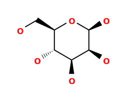 BMA
, MANMAN
, FUCFUC
), LA (IODIOD
), LB (IODIOD
), M (IODIOD
), MA (IODIOD
), MB (IODIOD
), MC (IODIOD
), N (IODIOD
), NA (IODIOD
), NB (IODIOD
), NC (IODIOD
), O (IODIOD
), OA (IODIOD
), OB (IODIOD
), OC (IODIOD
), P (IODIOD
), PA (IODIOD
), PB (IODIOD
), PC (IODIOD
), Q (IODIOD
), QA (IODIOD
), QB (IODIOD
), QC (IODIOD
), R (IODIOD
), RA (IODIOD
), RB (IODIOD
), RC (IODIOD
), S (IODIOD
), SA (IODIOD
), SB (IODIOD
), SC (IODIOD
), T (IODIOD
), TA (IODIOD
), TB (IODIOD
), TC (IODIOD
), U (IODIOD
), UA (IODIOD
), UB (IODIOD
), UC (IODIOD
), V (IODIOD
), VA (IODIOD
), VB (IODIOD
), VC (IODIOD
), W (IODIOD
), WA (IODIOD
), WB (IODIOD
), WC (IODIOD
), X (IODIOD
), XA (IODIOD
), XB (IODIOD
), XC (IODIOD
), Y (IODIOD
), YA (IODIOD
), YB (IODIOD
), YC (IODIOD
), Z (IODIOD
), ZA (IODIOD
), ZB (IODIOD
), ZC (IODIOD
) |

Each chain information
Showing 2 chains with heme of 6. Show all chains
| auth_asym_id | B |
|---|---|
| Residue indices (Uniprot) | 279-744 (279-745) |
| Missing residues | 316CYS-316CYS |
| Nonstandard amino acids |
 CSO |
| Uniprot ID | P05164 →UniprotKB |
| S-S bonds | 281:291,285:309,387:398,606:663,704:730 |
| label_asym_ids of heme | LC |
| Available structure | PDB and Modeled |
| RMSD of experimental residues after energy minimization | 0.10 Å |
| auth_asym_id | D |
|---|---|
| Residue indices (Uniprot) | 279-743 (279-745) |
| Missing residues | 316CYS-316CYS |
| Nonstandard amino acids |
CSO
|
| Uniprot ID | P05164 →UniprotKB |
| S-S bonds | 281:291,285:309,387:398,606:663,704:730 |
| label_asym_ids of heme | CB |
| Available structure | PDB and Modeled |
| RMSD of experimental residues after energy minimization | 0.11 Å |