
Number of data: 7660
Dataset: all data
| pdb_00003b99 | Crystal structure of zebrafish prostacyclin synthase (cytochrome P450 8A1) in complex with substrate analog U51605 | ||
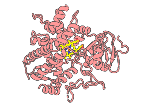 |
Experimental method | X-RAY DIFFRACTION |
|---|---|---|
| Structural data | available | |
| Non-standard amino acids | None | |
| Missing residues/ Structural gap | Exists / Exists | |
| Resolution | 2.5 Å | |
| Deposition date | 2007-11-03 |
| pdb_00003bcc | STIGMATELLIN AND ANTIMYCIN BOUND CYTOCHROME BC1 COMPLEX FROM CHICKEN | ||
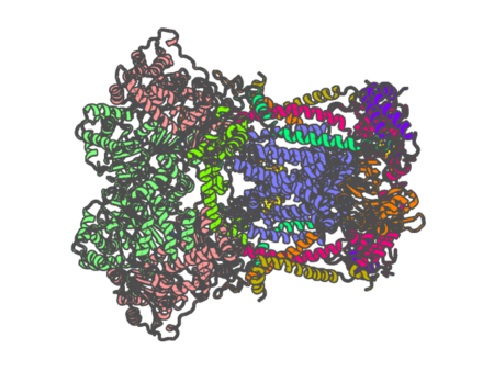 |
Experimental method | X-RAY DIFFRACTION |
|---|---|---|
| Structural data | available | |
| Non-standard amino acids | None | |
| Missing residues/ Structural gap | Exists / None | |
| Resolution | 3.7 Å | |
| Deposition date | 1998-03-23 |
| pdb_00003bcq | Crystal structure of oxy-hemoglobin from Brycon cephalus | ||
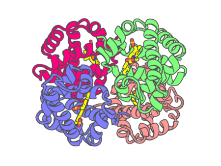 |
Experimental method | X-RAY DIFFRACTION |
|---|---|---|
| Structural data | available | |
| Non-standard amino acids | None | |
| Missing residues/ Structural gap | None / None | |
| Resolution | 2.4 Å | |
| Deposition date | 2007-11-13 |
| pdb_00003bdz | The Role of Asn 242 in P450cin | ||
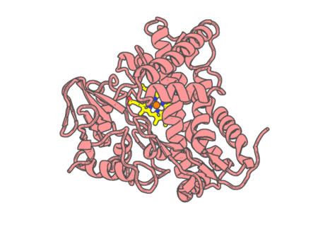 |
Experimental method | X-RAY DIFFRACTION |
|---|---|---|
| Structural data | available | |
| Non-standard amino acids | None | |
| Missing residues/ Structural gap | None / None | |
| Resolution | 2 Å | |
| Deposition date | 2007-11-15 |
| pdb_00003be0 | The Role of Asn 242 in P450cin | ||
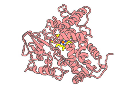 |
Experimental method | X-RAY DIFFRACTION |
|---|---|---|
| Structural data | available | |
| Non-standard amino acids | None | |
| Missing residues/ Structural gap | None / None | |
| Resolution | 3.05 Å | |
| Deposition date | 2007-11-15 |
| pdb_00003ben | Structure of N-(12-imidazolyl-dodecanoyl)-L-leucine inhibitor bound to the heme domain of Cytochrome P450-BM3 | ||
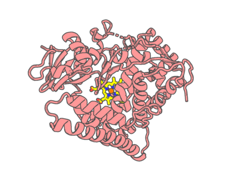 |
Experimental method | X-RAY DIFFRACTION |
|---|---|---|
| Structural data | available | |
| Non-standard amino acids | None | |
| Missing residues/ Structural gap | None / None | |
| Resolution | 1.65 Å | |
| Deposition date | 2007-11-19 |
| pdb_00003bj1 | met-Perch Hemoglobin at pH 5.7 | ||
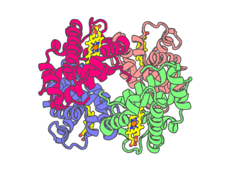 |
Experimental method | X-RAY DIFFRACTION |
|---|---|---|
| Structural data | available | |
| Non-standard amino acids | None | |
| Missing residues/ Structural gap | Exists / Exists | |
| Resolution | 1.9 Å | |
| Deposition date | 2007-12-02 |
| pdb_00003bj2 | met-Perch Hemoglobin at pH 6.3 | ||
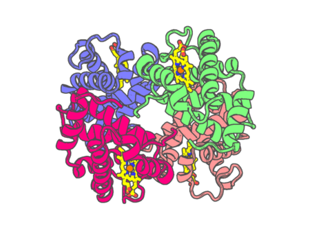 |
Experimental method | X-RAY DIFFRACTION |
|---|---|---|
| Structural data | available | |
| Non-standard amino acids | None | |
| Missing residues/ Structural gap | None / None | |
| Resolution | 2 Å | |
| Deposition date | 2007-12-02 |
| pdb_00003bj3 | met-Perch hemoglobin at pH 8.0 | ||
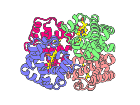 |
Experimental method | X-RAY DIFFRACTION |
|---|---|---|
| Structural data | available | |
| Non-standard amino acids | None | |
| Missing residues/ Structural gap | None / None | |
| Resolution | 2.1 Å | |
| Deposition date | 2007-12-02 |
| pdb_00003bk9 | H55A mutant of tryptophan 2,3-dioxygenase from Xanthomonas campestris | ||
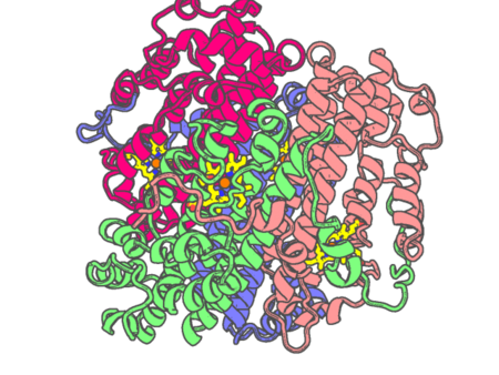 |
Experimental method | X-RAY DIFFRACTION |
|---|---|---|
| Structural data | available | |
| Non-standard amino acids | None | |
| Missing residues/ Structural gap | Exists / Exists | |
| Resolution | 2.15 Å | |
| Deposition date | 2007-12-06 |
| pdb_00003bkn | The structure of Mycobacterial bacterioferritin | ||
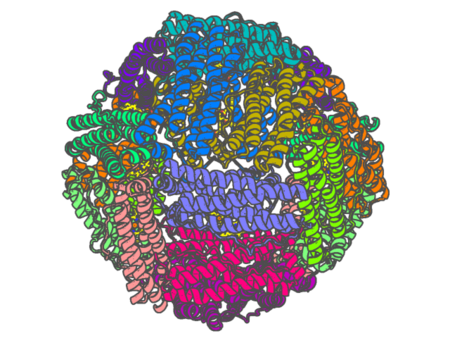 |
Experimental method | X-RAY DIFFRACTION |
|---|---|---|
| Structural data | available | |
| Non-standard amino acids | None | |
| Missing residues/ Structural gap | None / None | |
| Resolution | 2.72 Å | |
| Deposition date | 2007-12-07 |
| pdb_00003bnf | W. succinogenes NrfA Sulfite Complex | ||
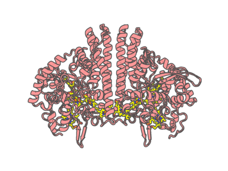 |
Experimental method | X-RAY DIFFRACTION |
|---|---|---|
| Structural data | available | |
| Non-standard amino acids | None | |
| Missing residues/ Structural gap | None / None | |
| Resolution | 1.7 Å | |
| Deposition date | 2007-12-14 |
| pdb_00003bng | W. succinogenes NrfA Y218F | ||
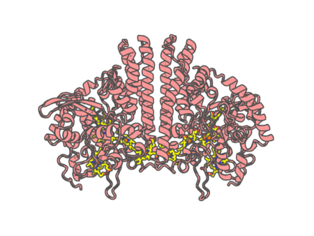 |
Experimental method | X-RAY DIFFRACTION |
|---|---|---|
| Structural data | available | |
| Non-standard amino acids | None | |
| Missing residues/ Structural gap | None / None | |
| Resolution | 1.5 Å | |
| Deposition date | 2007-12-14 |
| pdb_00003bnh | W. succinogenes NrfA Y218F Nitrite Complex | ||
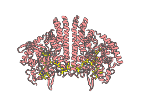 |
Experimental method | X-RAY DIFFRACTION |
|---|---|---|
| Structural data | available | |
| Non-standard amino acids | None | |
| Missing residues/ Structural gap | None / None | |
| Resolution | 1.75 Å | |
| Deposition date | 2007-12-14 |
| pdb_00003bnj | W. succinogenes NrfA Y218F Sulfite Complex | ||
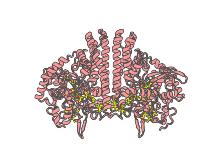 |
Experimental method | X-RAY DIFFRACTION |
|---|---|---|
| Structural data | available | |
| Non-standard amino acids | None | |
| Missing residues/ Structural gap | None / None | |
| Resolution | 1.3 Å | |
| Deposition date | 2007-12-14 |
| pdb_00003bom | Crystal structure of trout hemoglobin at 1.35 Angstrom resolution | ||
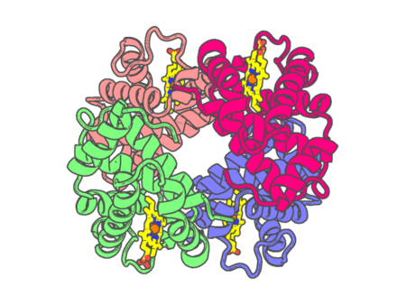 |
Experimental method | X-RAY DIFFRACTION |
|---|---|---|
| Structural data | available | |
| Non-standard amino acids | None | |
| Missing residues/ Structural gap | None / None | |
| Resolution | 1.35 Å | |
| Deposition date | 2007-12-17 |
| pdb_00003buj | Crystal Structure of CalO2 | ||
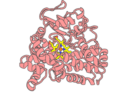 |
Experimental method | X-RAY DIFFRACTION |
|---|---|---|
| Structural data | available | |
| Non-standard amino acids | None | |
| Missing residues/ Structural gap | None / None | |
| Resolution | 2.47 Å | |
| Deposition date | 2008-01-02 |
| pdb_00003bvd | Structure of Surface-engineered Cytochrome ba3 Oxidase from Thermus thermophilus under Xenon Pressure, 100psi 5min | ||
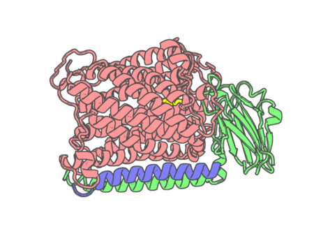 |
Experimental method | X-RAY DIFFRACTION |
|---|---|---|
| Structural data | available | |
| Non-standard amino acids | None | |
| Missing residues/ Structural gap | None / None | |
| Resolution | 3.37 Å | |
| Deposition date | 2008-01-07 |
| pdb_00003bxi | Structure of the complex of bovine lactoperoxidase with its catalyzed product hypothiocyanate ion at 2.3A resolution | ||
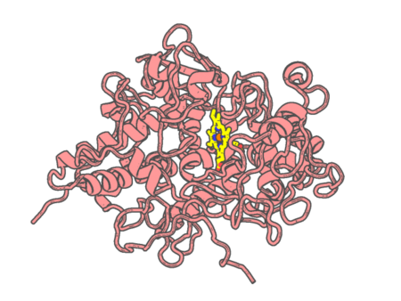 |
Experimental method | X-RAY DIFFRACTION |
|---|---|---|
| Structural data | available | |
| Non-standard amino acids | Included | |
| Missing residues/ Structural gap | None / None | |
| Resolution | 2.3 Å | |
| Deposition date | 2008-01-14 |
| pdb_00003bxu | PpcB, A Cytochrome c7 from Geobacter sulfurreducens | ||
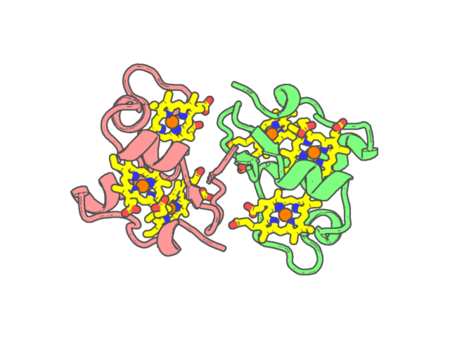 |
Experimental method | X-RAY DIFFRACTION |
|---|---|---|
| Structural data | available | |
| Non-standard amino acids | None | |
| Missing residues/ Structural gap | None / None | |
| Resolution | 1.35 Å | |
| Deposition date | 2008-01-14 |