
Number of data: 7660
Dataset: all data
| pdb_00005agk | Structure of rat neuronal nitric oxide synthase heme domain in complex with (2S)-2-Amino-5-(2-(methylsulfinyl)acetimidamido) pentanoic acid | ||
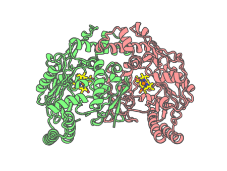 |
Experimental method | X-RAY DIFFRACTION |
|---|---|---|
| Structural data | available | |
| Non-standard amino acids | None | |
| Missing residues/ Structural gap | Exists / Exists | |
| Resolution | 2 Å | |
| Deposition date | 2015-02-02 |
| pdb_00005agl | Structure of rat neuronal nitric oxide synthase heme domain in complex with (S)-2-Amino-5-(2-(methylsulfonyl)acetimidamido)pentanoic acid | ||
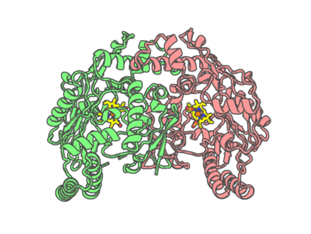 |
Experimental method | X-RAY DIFFRACTION |
|---|---|---|
| Structural data | available | |
| Non-standard amino acids | None | |
| Missing residues/ Structural gap | Exists / Exists | |
| Resolution | 1.94 Å | |
| Deposition date | 2015-02-02 |
| pdb_00005agm | Structure of rat neuronal nitric oxide synthase heme domain in complex with (S)-2-Amino-5-(2-oxoacetimidamido)pentanoic acid | ||
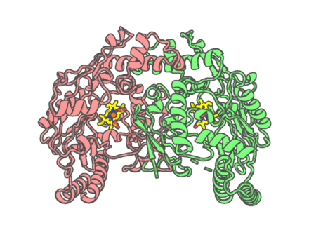 |
Experimental method | X-RAY DIFFRACTION |
|---|---|---|
| Structural data | available | |
| Non-standard amino acids | None | |
| Missing residues/ Structural gap | Exists / Exists | |
| Resolution | 1.84 Å | |
| Deposition date | 2015-02-02 |
| pdb_00005agn | Structure of rat neuronal nitric oxide synthase heme domain in complex with (S)-2-Amino-5-(2-hydroxyacetimidamido)pentanoic acid | ||
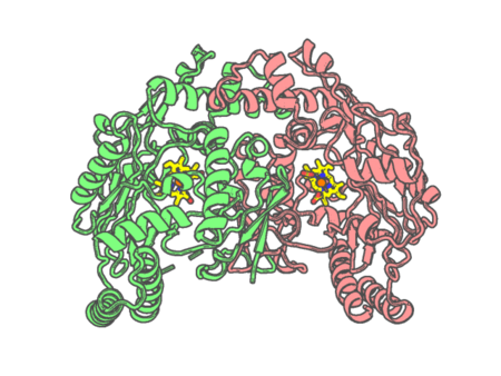 |
Experimental method | X-RAY DIFFRACTION |
|---|---|---|
| Structural data | available | |
| Non-standard amino acids | None | |
| Missing residues/ Structural gap | Exists / Exists | |
| Resolution | 1.95 Å | |
| Deposition date | 2015-02-02 |
| pdb_00005ago | Structure of rat neuronal nitric oxide synthase heme domain in complex with (S)-2-Amino-5-(2-mercaptoacetimidamido)pentanoic acid | ||
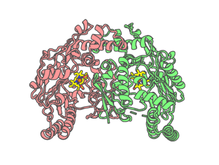 |
Experimental method | X-RAY DIFFRACTION |
|---|---|---|
| Structural data | available | |
| Non-standard amino acids | None | |
| Missing residues/ Structural gap | Exists / Exists | |
| Resolution | 1.902 Å | |
| Deposition date | 2015-02-02 |
| pdb_00005agp | Structure of rat neuronal nitric oxide synthase heme domain in complex with (S)-2-Amino-5-(2-mercaptoacetimidamido)pentanoic acid | ||
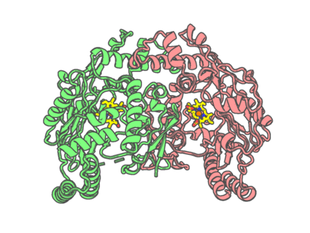 |
Experimental method | X-RAY DIFFRACTION |
|---|---|---|
| Structural data | available | |
| Non-standard amino acids | None | |
| Missing residues/ Structural gap | Exists / Exists | |
| Resolution | 2.1 Å | |
| Deposition date | 2015-02-02 |
| pdb_00005ajr | Sterol 14-alpha demethylase (CYP51) from Trypanosoma cruzi in complex with the 1-tetrazole derivative VT-1161 ((R)-2-(2,4-Difluorophenyl)-1, 1-difluoro-3-(1H-tetrazol-1-yl)-1-(5-(4-(2,2,2-trifluoroethoxy)phenyl) pyridin-2-yl)propan-2-ol) | ||
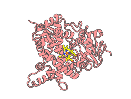 |
Experimental method | X-RAY DIFFRACTION |
|---|---|---|
| Structural data | available | |
| Non-standard amino acids | None | |
| Missing residues/ Structural gap | Exists / Exists | |
| Resolution | 2.75 Å | |
| Deposition date | 2015-02-26 |
| pdb_00005al9 | Structure of Leishmania major peroxidase D211R mutant (high res) | ||
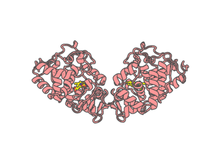 |
Experimental method | X-RAY DIFFRACTION |
|---|---|---|
| Structural data | available | |
| Non-standard amino acids | None | |
| Missing residues/ Structural gap | None / None | |
| Resolution | 1.37 Å | |
| Deposition date | 2015-03-07 |
| pdb_00005ala | Structure of Leishmania major peroxidase D211R mutant (low res) | ||
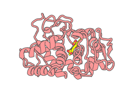 |
Experimental method | X-RAY DIFFRACTION |
|---|---|---|
| Structural data | available | |
| Non-standard amino acids | None | |
| Missing residues/ Structural gap | None / None | |
| Resolution | 2.73 Å | |
| Deposition date | 2015-03-07 |
| pdb_00005amm | Structure of Leishmania major peroxidase D211N mutant | ||
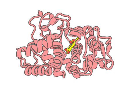 |
Experimental method | X-RAY DIFFRACTION |
|---|---|---|
| Structural data | available | |
| Non-standard amino acids | None | |
| Missing residues/ Structural gap | None / None | |
| Resolution | 2.09 Å | |
| Deposition date | 2015-03-11 |
| pdb_00005aog | Structure of Sorghum peroxidase | ||
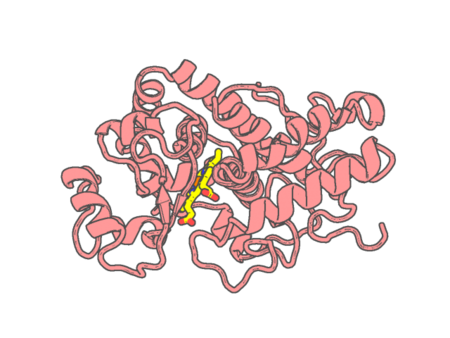 |
Experimental method | X-RAY DIFFRACTION |
|---|---|---|
| Structural data | available | |
| Non-standard amino acids | None | |
| Missing residues/ Structural gap | None / None | |
| Resolution | 1.27 Å | |
| Deposition date | 2015-09-10 |
| pdb_00005aur | Hydrogenobacter thermophilus cytochrome c552 dimer formed by domain swapping at N-terminal region | ||
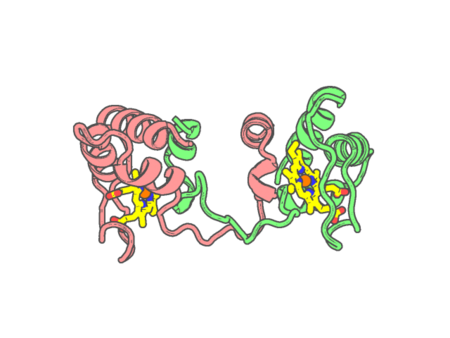 |
Experimental method | X-RAY DIFFRACTION |
|---|---|---|
| Structural data | available | |
| Non-standard amino acids | None | |
| Missing residues/ Structural gap | None / None | |
| Resolution | 1.26 Å | |
| Deposition date | 2015-06-08 |
| pdb_00005aus | Hydrogenobacter thermophilus cytochrome c552 dimer formed by domain swapping at C-terminal region | ||
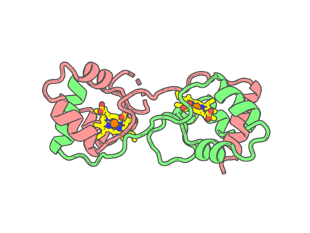 |
Experimental method | X-RAY DIFFRACTION |
|---|---|---|
| Structural data | available | |
| Non-standard amino acids | None | |
| Missing residues/ Structural gap | Exists / Exists | |
| Resolution | 1.3 Å | |
| Deposition date | 2015-06-08 |
| pdb_00005awi | Domain-swapped cytochrome cb562 dimer | ||
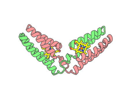 |
Experimental method | X-RAY DIFFRACTION |
|---|---|---|
| Structural data | available | |
| Non-standard amino acids | None | |
| Missing residues/ Structural gap | None / None | |
| Resolution | 1.85 Å | |
| Deposition date | 2015-07-03 |
| pdb_00005az3 | Crystal structure of heme binding protein HmuT | ||
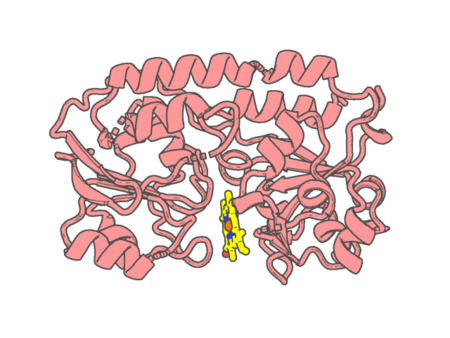 |
Experimental method | X-RAY DIFFRACTION |
|---|---|---|
| Structural data | available | |
| Non-standard amino acids | None | |
| Missing residues/ Structural gap | None / None | |
| Resolution | 1.423 Å | |
| Deposition date | 2015-09-18 |
| pdb_00005b1a | Bovine heart cytochrome c oxidase in the fully oxidized state at 1.5 angstrom resolution | ||
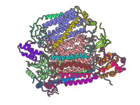 |
Experimental method | X-RAY DIFFRACTION |
|---|---|---|
| Structural data | available | |
| Non-standard amino acids | Included | |
| Missing residues/ Structural gap | None / None | |
| Resolution | 1.5 Å | |
| Deposition date | 2015-12-01 |
| pdb_00005b1b | Bovine heart cytochrome c oxidase in the fully reduced state at 1.6 angstrom resolution | ||
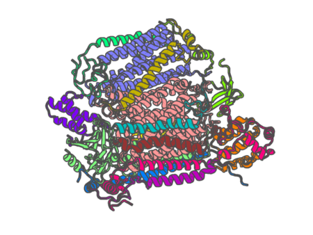 |
Experimental method | X-RAY DIFFRACTION |
|---|---|---|
| Structural data | available | |
| Non-standard amino acids | Included | |
| Missing residues/ Structural gap | None / None | |
| Resolution | 1.6 Å | |
| Deposition date | 2015-12-01 |
| pdb_00005b2u | Crystal Structure of P450BM3 with N-perfluorohexanoyl -L-tryptophan | ||
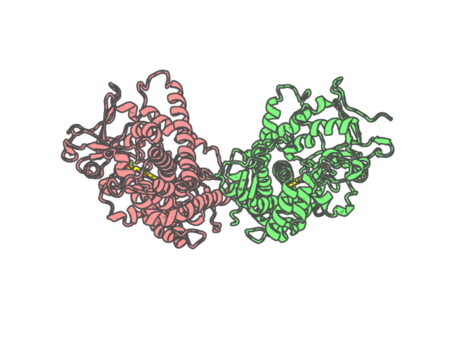 |
Experimental method | X-RAY DIFFRACTION |
|---|---|---|
| Structural data | available | |
| Non-standard amino acids | None | |
| Missing residues/ Structural gap | None / None | |
| Resolution | 1.9 Å | |
| Deposition date | 2016-02-03 |
| pdb_00005b2v | Crystal Structure of P450BM3 with N-perfluorohexanoyl-L-tryptophan | ||
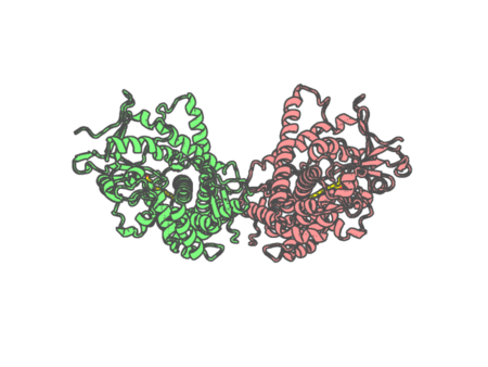 |
Experimental method | X-RAY DIFFRACTION |
|---|---|---|
| Structural data | available | |
| Non-standard amino acids | None | |
| Missing residues/ Structural gap | None / None | |
| Resolution | 2.3 Å | |
| Deposition date | 2016-02-03 |
| pdb_00005b2w | Crystal Structure of P450BM3 with N-perfluorododecanoyl-L-tryptophan | ||
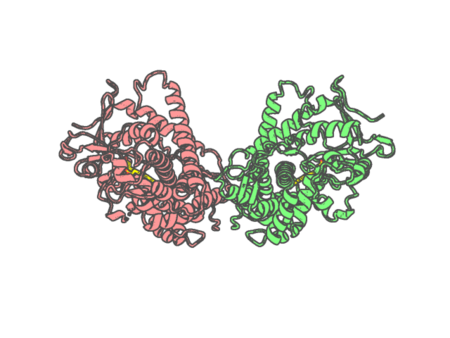 |
Experimental method | X-RAY DIFFRACTION |
|---|---|---|
| Structural data | available | |
| Non-standard amino acids | None | |
| Missing residues/ Structural gap | None / None | |
| Resolution | 1.65 Å | |
| Deposition date | 2016-02-03 |