Number of data: 7660
Dataset: all data
| pdb_00006wr1 |
Human steroidogenic cytochrome P450 17A1 mutant N52Y with inhibitor abiraterone
|
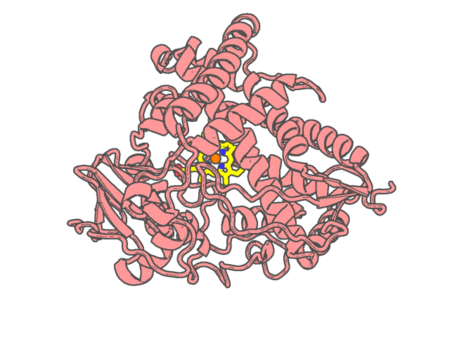 |
Experimental method |
X-RAY DIFFRACTION |
| Structural data |
available |
| Non-standard amino acids |
None |
| Missing residues/ Structural gap |
Exists / Exists |
| Resolution |
1.85 Å |
| Deposition date |
2020-04-29 |
| pdb_00006wti |
The Cryo-EM structure of the ubiquinol oxidase from Escherichia coli
|
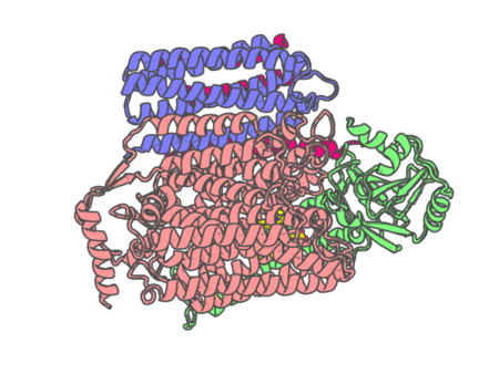 |
Experimental method |
ELECTRON MICROSCOPY |
| Structural data |
available |
| Non-standard amino acids |
None |
| Missing residues/ Structural gap |
None / None |
| Resolution |
None |
| Deposition date |
2020-05-02 |
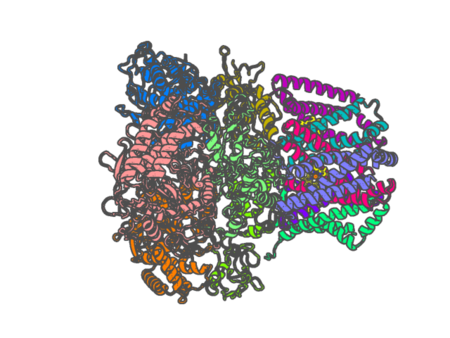 |
Experimental method |
ELECTRON MICROSCOPY |
| Structural data |
unavailable |
| Non-standard amino acids |
None |
| Missing residues/ Structural gap |
Exists / Exists |
| Resolution |
None |
| Deposition date |
2020-05-04 |
| pdb_00006ww0 |
Human steroidogenic cytochrome P450 17A1 with 3-keto-5alpha-abiraterone analog
|
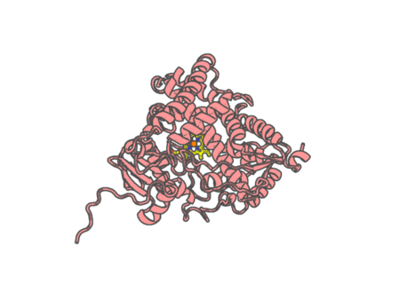 |
Experimental method |
X-RAY DIFFRACTION |
| Structural data |
available |
| Non-standard amino acids |
None |
| Missing residues/ Structural gap |
Exists / Exists |
| Resolution |
2.01 Å |
| Deposition date |
2020-05-07 |
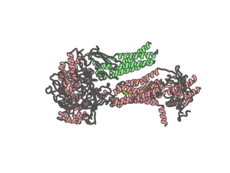
| pdb_00006wxr |
CryoEM structure of mouse DUOX1-DUOXA1 complex in the absence of NADPH
|
 |
Experimental method |
ELECTRON MICROSCOPY |
| Structural data |
unavailable |
| Non-standard amino acids |
None |
| Missing residues/ Structural gap |
Exists / Exists |
| Resolution |
None |
| Deposition date |
2020-05-11 |
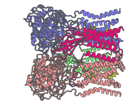
| pdb_00006wxu |
CryoEM structure of mouse DUOX1-DUOXA1 complex in the dimer-of-dimer state
|
 |
Experimental method |
ELECTRON MICROSCOPY |
| Structural data |
unavailable |
| Non-standard amino acids |
None |
| Missing residues/ Structural gap |
Exists / Exists |
| Resolution |
None |
| Deposition date |
2020-05-12 |
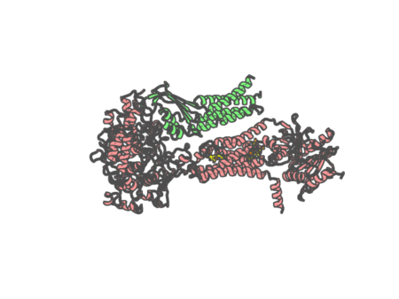
| pdb_00006wxv |
CryoEM structure of mouse DUOX1-DUOXA1 complex in the presence of NADPH
|
 |
Experimental method |
ELECTRON MICROSCOPY |
| Structural data |
unavailable |
| Non-standard amino acids |
None |
| Missing residues/ Structural gap |
Exists / Exists |
| Resolution |
None |
| Deposition date |
2020-05-12 |
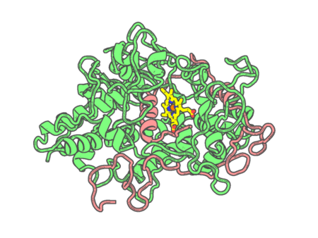
| pdb_00006wxz |
CRYSTAL STRUCTURE OF MYELOPEROXIDASE SUBFORM C (MPO) COMPLEX WITH Compound-29 A.K.A 7-(1,2-DIPHENYLETHYL)-1H-[1,2,3]TRIAZOLO[4,5-B]PYRIDIN-5-AMINE
|
 |
Experimental method |
X-RAY DIFFRACTION |
| Structural data |
available |
| Non-standard amino acids |
Included |
| Missing residues/ Structural gap |
None / None |
| Resolution |
2.226 Å |
| Deposition date |
2020-05-12 |
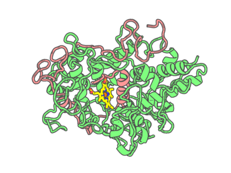
| pdb_00006wy0 |
CRYSTAL STRUCTURE OF MYELOPEROXIDASE SUBFORM C (MPO) COMPLEX WITH Compound-40 A.K.A 7-[(1R)-1-phenyl-3-{[(1r,4r)-4-phenylcyclohexyl]amino}propyl]-3H-[1,2,3]triazolo[4,5-b]pyridin-5-amine
|
 |
Experimental method |
X-RAY DIFFRACTION |
| Structural data |
available |
| Non-standard amino acids |
Included |
| Missing residues/ Structural gap |
None / None |
| Resolution |
2.799 Å |
| Deposition date |
2020-05-12 |
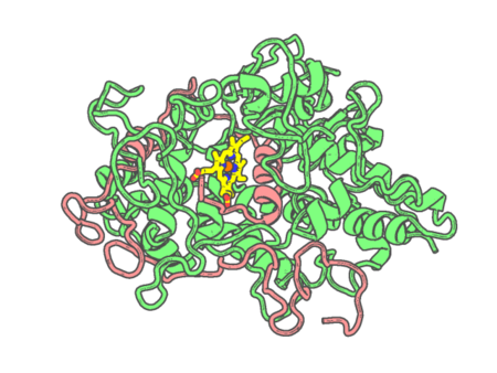
| pdb_00006wy5 |
CRYSTAL STRUCTURE OF MYELOPEROXIDASE SUBFORM C (MPO) COMPLEX WITH Compound-37 A.K.A 7-(1-phenyl-3-(((1S,3S)-3-phenyl-2,3-dihydro-1H-inden-1-yl)amino)propyl)-1H-[1,2,3]triazolo[4,5-b]pyridin-5-amine
|
 |
Experimental method |
X-RAY DIFFRACTION |
| Structural data |
available |
| Non-standard amino acids |
Included |
| Missing residues/ Structural gap |
None / None |
| Resolution |
2.898 Å |
| Deposition date |
2020-05-12 |
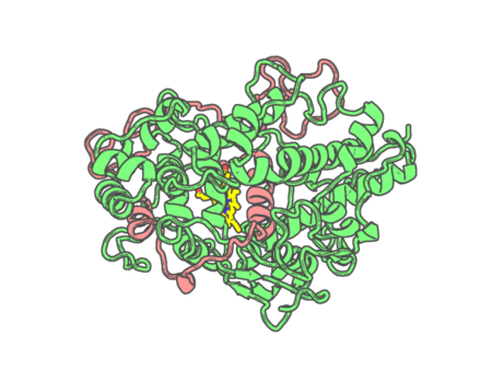
| pdb_00006wy7 |
CRYSTAL STRUCTURE OF MYELOPEROXIDASE SUBFORM C (MPO) COMPLEX WITH Compound-41 A.K.A 7-[1-phenyl-3-({4-phenylbicyclo[2.2.2]octan-1-yl}amino)propyl]-3H-[1,2,3]triazolo[4,5-b]pyridin-5-amine
|
 |
Experimental method |
X-RAY DIFFRACTION |
| Structural data |
available |
| Non-standard amino acids |
Included |
| Missing residues/ Structural gap |
None / None |
| Resolution |
2.089 Å |
| Deposition date |
2020-05-12 |
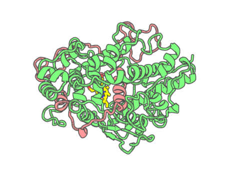
| pdb_00006wyd |
CRYSTAL STRUCTURE OF MYELOPEROXIDASE SUBFORM C (MPO) COMPLEX WITH Compound-12 (AKA; 7-benzyl-1H-[1,2,3]triazolo[4,5-b]pyrid
|
 |
Experimental method |
X-RAY DIFFRACTION |
| Structural data |
available |
| Non-standard amino acids |
None |
| Missing residues/ Structural gap |
None / None |
| Resolution |
2.55 Å |
| Deposition date |
2020-05-12 |
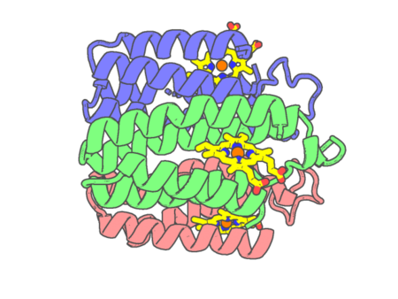 |
Experimental method |
X-RAY DIFFRACTION |
| Structural data |
available |
| Non-standard amino acids |
None |
| Missing residues/ Structural gap |
None / None |
| Resolution |
1.76 Å |
| Deposition date |
2020-05-13 |
| pdb_00006wz0 |
Fe-bound structure of an engineered metal-dependent protein trimer, TriCyt2
|
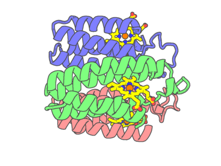 |
Experimental method |
X-RAY DIFFRACTION |
| Structural data |
available |
| Non-standard amino acids |
None |
| Missing residues/ Structural gap |
None / None |
| Resolution |
1.7 Å |
| Deposition date |
2020-05-13 |
| pdb_00006wz1 |
Mn-bound structure of an engineered protein trimer, TriCyt3
|
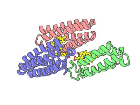 |
Experimental method |
X-RAY DIFFRACTION |
| Structural data |
available |
| Non-standard amino acids |
None |
| Missing residues/ Structural gap |
None / None |
| Resolution |
1.999 Å |
| Deposition date |
2020-05-13 |
| pdb_00006wz2 |
Co-bound structure of an engineered protein trimer, TriCyt3
|
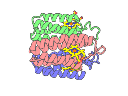 |
Experimental method |
X-RAY DIFFRACTION |
| Structural data |
available |
| Non-standard amino acids |
None |
| Missing residues/ Structural gap |
None / None |
| Resolution |
2.0006 Å |
| Deposition date |
2020-05-13 |
| pdb_00006wz3 |
Cu-bound structure of the engineered protein trimer, TriCyt3
|
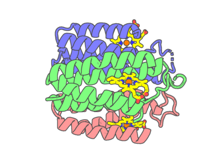 |
Experimental method |
X-RAY DIFFRACTION |
| Structural data |
available |
| Non-standard amino acids |
None |
| Missing residues/ Structural gap |
None / None |
| Resolution |
1.8 Å |
| Deposition date |
2020-05-13 |
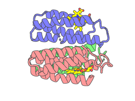 |
Experimental method |
X-RAY DIFFRACTION |
| Structural data |
available |
| Non-standard amino acids |
None |
| Missing residues/ Structural gap |
None / None |
| Resolution |
2.3 Å |
| Deposition date |
2020-05-13 |
| pdb_00006wza |
Ni-bound structure of an engineered metal-dependent protein trimer, TriCyt1
|
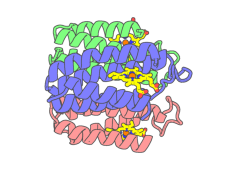 |
Experimental method |
X-RAY DIFFRACTION |
| Structural data |
available |
| Non-standard amino acids |
None |
| Missing residues/ Structural gap |
None / None |
| Resolution |
2.5 Å |
| Deposition date |
2020-05-13 |
| pdb_00006wzc |
Ni-bound structure of an engineered protein trimer, TriCyt3
|
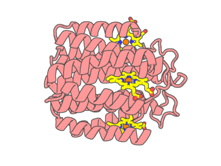 |
Experimental method |
X-RAY DIFFRACTION |
| Structural data |
available |
| Non-standard amino acids |
None |
| Missing residues/ Structural gap |
None / None |
| Resolution |
2.195 Å |
| Deposition date |
2020-05-13 |
