
Number of data: 7660
Dataset: all data
| pdb_00007dgl | The Ni-bound dimeric structure of K78H/G80A/H82A myoglobin | ||
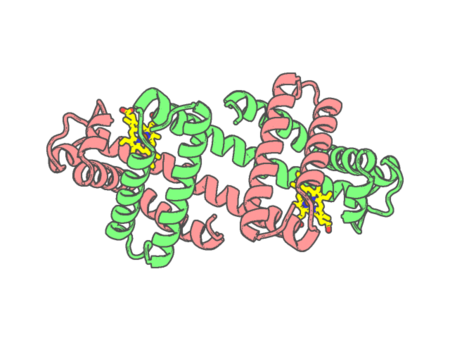 |
Experimental method | X-RAY DIFFRACTION |
|---|---|---|
| Structural data | available | |
| Non-standard amino acids | None | |
| Missing residues/ Structural gap | None / None | |
| Resolution | 1.91 Å | |
| Deposition date | 2020-11-12 |
| pdb_00007dgm | The dimeric structure of K79H/G80A/H81A myoglobin | ||
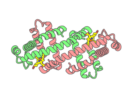 |
Experimental method | X-RAY DIFFRACTION |
|---|---|---|
| Structural data | available | |
| Non-standard amino acids | None | |
| Missing residues/ Structural gap | None / None | |
| Resolution | 1.62 Å | |
| Deposition date | 2020-11-12 |
| pdb_00007dgn | The Co-bound dimeric structure of K79H/G80A/H81A myoglobin | ||
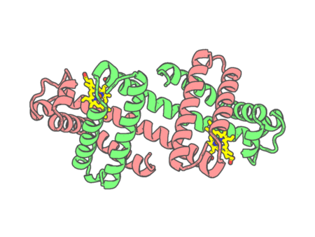 |
Experimental method | X-RAY DIFFRACTION |
|---|---|---|
| Structural data | available | |
| Non-standard amino acids | None | |
| Missing residues/ Structural gap | None / None | |
| Resolution | 2.35 Å | |
| Deposition date | 2020-11-12 |
| pdb_00007dgo | The Zn-bound dimeric structure of K79H/G80A/H81A myoglobin | ||
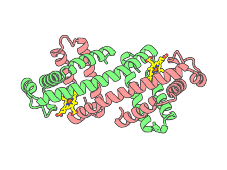 |
Experimental method | X-RAY DIFFRACTION |
|---|---|---|
| Structural data | available | |
| Non-standard amino acids | None | |
| Missing residues/ Structural gap | None / None | |
| Resolution | 2 Å | |
| Deposition date | 2020-11-12 |
| pdb_00007dgq | Activity optimized supercomplex state1 | ||
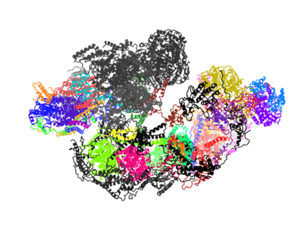 |
Experimental method | ELECTRON MICROSCOPY |
|---|---|---|
| Structural data | available | |
| Non-standard amino acids | None | |
| Missing residues/ Structural gap | Exists / Exists | |
| Resolution | None | |
| Deposition date | 2020-11-12 |
| pdb_00007dgr | Activity optimized supercomplex state2 | ||
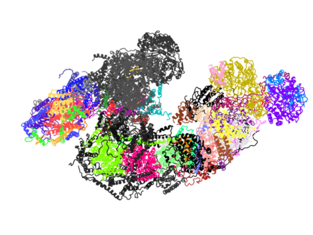 |
Experimental method | ELECTRON MICROSCOPY |
|---|---|---|
| Structural data | available | |
| Non-standard amino acids | None | |
| Missing residues/ Structural gap | Exists / Exists | |
| Resolution | None | |
| Deposition date | 2020-11-12 |
| pdb_00007dgs | Activity optimized supercomplex state3 | ||
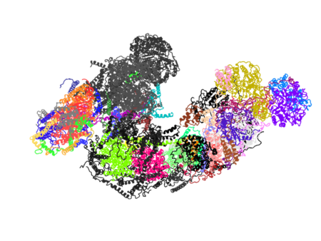 |
Experimental method | ELECTRON MICROSCOPY |
|---|---|---|
| Structural data | available | |
| Non-standard amino acids | None | |
| Missing residues/ Structural gap | Exists / Exists | |
| Resolution | None | |
| Deposition date | 2020-11-12 |
| pdb_00007di3 | Cytochrome P450 (CYP105D18) W.T. | ||
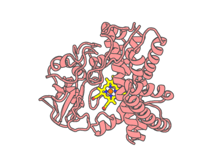 |
Experimental method | X-RAY DIFFRACTION |
|---|---|---|
| Structural data | available | |
| Non-standard amino acids | None | |
| Missing residues/ Structural gap | Exists / Exists | |
| Resolution | 1.69 Å | |
| Deposition date | 2020-11-18 |
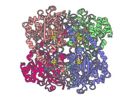
| pdb_00007di8 | Electron crystallographic structure of Catalase using a direct electron detector at 300 kV | ||
 |
Experimental method | ELECTRON CRYSTALLOGRAPHY |
|---|---|---|
| Structural data | available | |
| Non-standard amino acids | None | |
| Missing residues/ Structural gap | None / None | |
| Resolution | 3.2 Å | |
| Deposition date | 2020-11-18 |
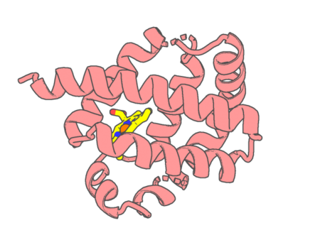
| pdb_00007dih | Crystal structure of Thermoglobin Y29F mutant in complex with imidazole | ||
 |
Experimental method | X-RAY DIFFRACTION |
|---|---|---|
| Structural data | available | |
| Non-standard amino acids | None | |
| Missing residues/ Structural gap | None / None | |
| Resolution | 1.5 Å | |
| Deposition date | 2020-11-19 |
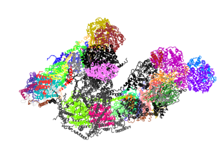
| pdb_00007dkf | Activity optimized supercomplex state4 | ||
 |
Experimental method | ELECTRON MICROSCOPY |
|---|---|---|
| Structural data | available | |
| Non-standard amino acids | None | |
| Missing residues/ Structural gap | Exists / Exists | |
| Resolution | None | |
| Deposition date | 2020-11-24 |
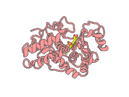
| pdb_00007dlh | Crystallization of Cationic Peroxidase from Proso Millet and Identification of Its Phosphatase Active Sites | ||
 |
Experimental method | X-RAY DIFFRACTION |
|---|---|---|
| Structural data | available | |
| Non-standard amino acids | None | |
| Missing residues/ Structural gap | None / None | |
| Resolution | 1.789 Å | |
| Deposition date | 2020-11-27 |
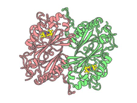
| pdb_00007dlk | Crystal Structure of veratryl alcohol bound Dye Decolorizing peroxidase from Bacillus subtilis | ||
 |
Experimental method | X-RAY DIFFRACTION |
|---|---|---|
| Structural data | available | |
| Non-standard amino acids | None | |
| Missing residues/ Structural gap | None / None | |
| Resolution | 2.1 Å | |
| Deposition date | 2020-11-27 |
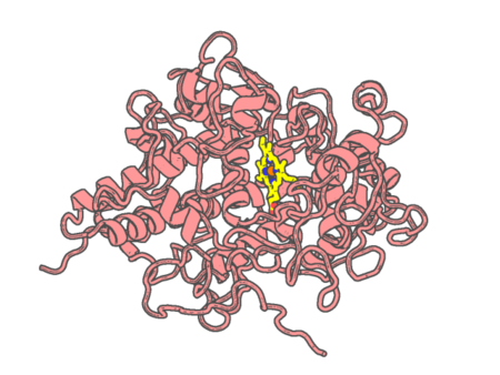
| pdb_00007dlq | CRYSTAL STRUCTURE OF THE COMPLEX OF LACTOPEROXIDASE WITH HYDROGEN PEROXIDE AT 1.77A RESOLUTION | ||
 |
Experimental method | X-RAY DIFFRACTION |
|---|---|---|
| Structural data | available | |
| Non-standard amino acids | None | |
| Missing residues/ Structural gap | None / None | |
| Resolution | 1.773 Å | |
| Deposition date | 2020-11-29 |
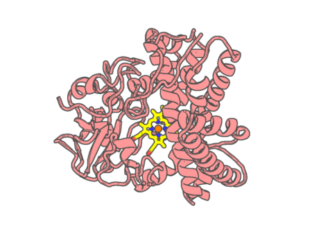
| pdb_00007dls | Cytochrome P450 (CYP105D18) complex with papaverine | ||
 |
Experimental method | X-RAY DIFFRACTION |
|---|---|---|
| Structural data | available | |
| Non-standard amino acids | None | |
| Missing residues/ Structural gap | Exists / Exists | |
| Resolution | 2.06 Å | |
| Deposition date | 2020-11-30 |
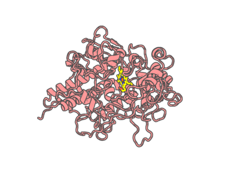
| pdb_00007dmr | Crystal structure of potassium induced heme modification in yak lactoperoxidase at 2.20 A resolution | ||
 |
Experimental method | X-RAY DIFFRACTION |
|---|---|---|
| Structural data | available | |
| Non-standard amino acids | None | |
| Missing residues/ Structural gap | None / None | |
| Resolution | 2.2 Å | |
| Deposition date | 2020-12-06 |
| pdb_00007dn6 | Crystal structure of bovine lactoperoxidase with hydrogen peroxide trapped between heme iron and his109 at 1.69 A resolution | ||
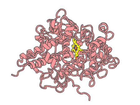 |
Experimental method | X-RAY DIFFRACTION |
|---|---|---|
| Structural data | available | |
| Non-standard amino acids | None | |
| Missing residues/ Structural gap | None / None | |
| Resolution | 1.696 Å | |
| Deposition date | 2020-12-08 |
| pdb_00007dn7 | Crystal structure of ternary complexes of lactoperoxidase with hydrogen peroxide at 1.70 A resolution | ||
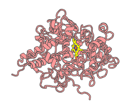 |
Experimental method | X-RAY DIFFRACTION |
|---|---|---|
| Structural data | available | |
| Non-standard amino acids | None | |
| Missing residues/ Structural gap | None / None | |
| Resolution | 1.7 Å | |
| Deposition date | 2020-12-09 |
| pdb_00007dnz | Cryo-EM structure of the human ABCB6 (Hemin and GSH-bound) | ||
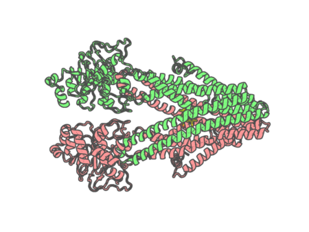 |
Experimental method | ELECTRON MICROSCOPY |
|---|---|---|
| Structural data | available | |
| Non-standard amino acids | None | |
| Missing residues/ Structural gap | None / None | |
| Resolution | None | |
| Deposition date | 2020-12-11 |
| pdb_00007dvo | Structure of Reaction Intermediate of Cytochrome P450 NO Reductase (P450nor) Determined by XFEL | ||
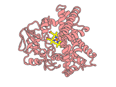 |
Experimental method | X-RAY DIFFRACTION |
|---|---|---|
| Structural data | available | |
| Non-standard amino acids | None | |
| Missing residues/ Structural gap | None / None | |
| Resolution | 1.8 Å | |
| Deposition date | 2021-01-14 |