
Number of data: 3440
Dataset: (without mutations)
| pdb_00005gkn | Catalase structure determined by electron crystallography of thin 3D crystals | ||
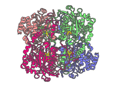 |
Experimental method | ELECTRON CRYSTALLOGRAPHY |
|---|---|---|
| Structural data | available | |
| Non-standard amino acids | None | |
| Missing residues/ Structural gap | None / None | |
| Resolution | 3.2 Å | |
| Deposition date | 2016-07-04 |
| pdb_00005gwe | cytochrome P450 CREJ | ||
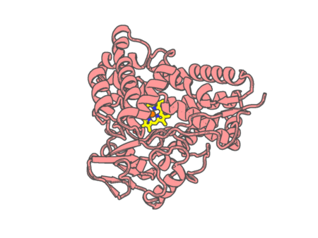 |
Experimental method | X-RAY DIFFRACTION |
|---|---|---|
| Structural data | available | |
| Non-standard amino acids | None | |
| Missing residues/ Structural gap | None / None | |
| Resolution | 2 Å | |
| Deposition date | 2016-09-11 |
| pdb_00005gyr | Tetrameric Allochromatium vinosum cytochrome c' | ||
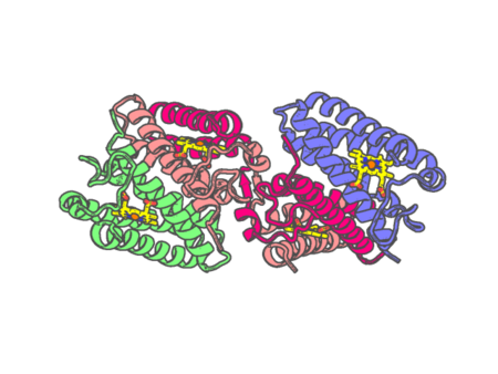 |
Experimental method | X-RAY DIFFRACTION |
|---|---|---|
| Structural data | available | |
| Non-standard amino acids | None | |
| Missing residues/ Structural gap | None / None | |
| Resolution | 1.6 Å | |
| Deposition date | 2016-09-23 |
| pdb_00005h1z | CYP153D17 from Sphingomonas sp. PAMC 26605 | ||
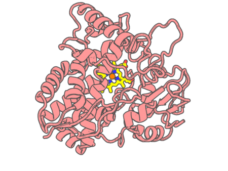 |
Experimental method | X-RAY DIFFRACTION |
|---|---|---|
| Structural data | available | |
| Non-standard amino acids | None | |
| Missing residues/ Structural gap | None / None | |
| Resolution | 3.1 Å | |
| Deposition date | 2016-10-13 |
| pdb_00005h2f | Crystal structure of the PsbM-deletion mutant of photosystem II | ||
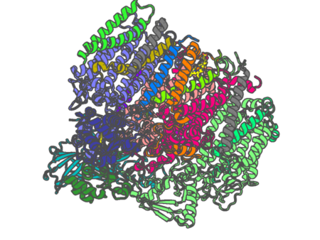 |
Experimental method | X-RAY DIFFRACTION |
|---|---|---|
| Structural data | available | |
| Non-standard amino acids | Included | |
| Missing residues/ Structural gap | None / None | |
| Resolution | 2.2 Å | |
| Deposition date | 2016-10-15 |
| pdb_00005hdi | Structural characterization of CYP144A1, a Mycobacterium tuberculosis cytochrome P450 | ||
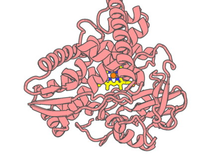 |
Experimental method | X-RAY DIFFRACTION |
|---|---|---|
| Structural data | available | |
| Non-standard amino acids | None | |
| Missing residues/ Structural gap | Exists / Exists | |
| Resolution | 1.54 Å | |
| Deposition date | 2016-01-05 |
| pdb_00005hh3 | OxyA from Actinoplanes teichomyceticus | ||
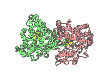 |
Experimental method | X-RAY DIFFRACTION |
|---|---|---|
| Structural data | available | |
| Non-standard amino acids | None | |
| Missing residues/ Structural gap | Exists / Exists | |
| Resolution | 2.1 Å | |
| Deposition date | 2016-01-09 |
| pdb_00005hiw | Sorangium cellulosum So Ce56 cytochrome P450 260B1 | ||
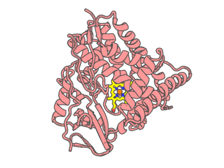 |
Experimental method | X-RAY DIFFRACTION |
|---|---|---|
| Structural data | available | |
| Non-standard amino acids | None | |
| Missing residues/ Structural gap | Exists / Exists | |
| Resolution | 1.85 Å | |
| Deposition date | 2016-01-12 |
| pdb_00005hs1 | Saccharomyces cerevisiae CYP51 (Lanosterol 14-alpha demethylase) complexed with Voriconazole | ||
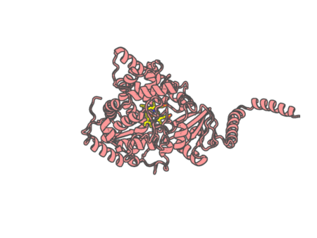 |
Experimental method | X-RAY DIFFRACTION |
|---|---|---|
| Structural data | available | |
| Non-standard amino acids | None | |
| Missing residues/ Structural gap | None / None | |
| Resolution | 2.1 Å | |
| Deposition date | 2016-01-24 |
| pdb_00005hy8 | Glycation restrains allosteric transition in hemoglobin: The molecular basis of oxidative stress under hyperglycemic conditions in diabetes | ||
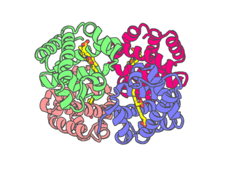 |
Experimental method | X-RAY DIFFRACTION |
|---|---|---|
| Structural data | available | |
| Non-standard amino acids | None | |
| Missing residues/ Structural gap | None / None | |
| Resolution | 2.3 Å | |
| Deposition date | 2016-02-01 |
| pdb_00005ibd | Crystal structure Mycobacterium tuberculosis CYP121 in complex with inhibitor fragment 24a | ||
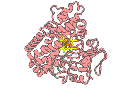 |
Experimental method | X-RAY DIFFRACTION |
|---|---|---|
| Structural data | available | |
| Non-standard amino acids | None | |
| Missing residues/ Structural gap | None / None | |
| Resolution | 1.77 Å | |
| Deposition date | 2016-02-22 |
| pdb_00005ibe | Crystal structure Mycobacterium tuberculosis CYP121 in complex with inhibitor fragment 25a | ||
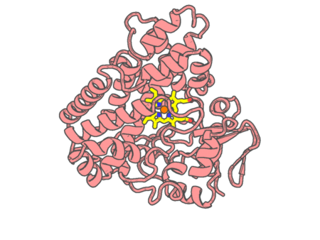 |
Experimental method | X-RAY DIFFRACTION |
|---|---|---|
| Structural data | available | |
| Non-standard amino acids | None | |
| Missing residues/ Structural gap | None / None | |
| Resolution | 1.624 Å | |
| Deposition date | 2016-02-22 |
| pdb_00005ibf | Crystal structure Mycobacterium tuberculosis CYP121 in complex with inhibitor fragment 19a | ||
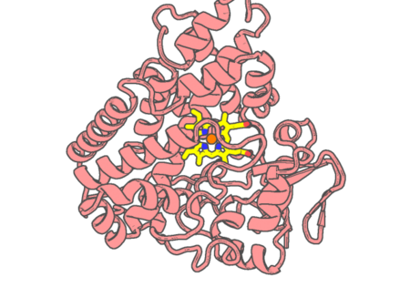 |
Experimental method | X-RAY DIFFRACTION |
|---|---|---|
| Structural data | available | |
| Non-standard amino acids | None | |
| Missing residues/ Structural gap | None / None | |
| Resolution | 1.7 Å | |
| Deposition date | 2016-02-22 |
| pdb_00005ibg | Crystal structure Mycobacterium tuberculosis CYP121 in complex with inhibitor fragment 25b | ||
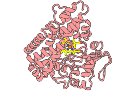 |
Experimental method | X-RAY DIFFRACTION |
|---|---|---|
| Structural data | available | |
| Non-standard amino acids | None | |
| Missing residues/ Structural gap | None / None | |
| Resolution | 2.1 Å | |
| Deposition date | 2016-02-22 |
| pdb_00005ibh | Crystal structure Mycobacterium tuberculosis CYP121 in complex with inhibitor fragment 26h | ||
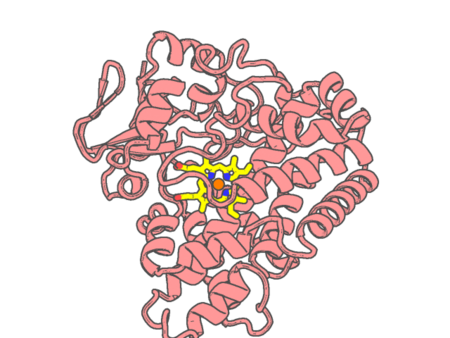 |
Experimental method | X-RAY DIFFRACTION |
|---|---|---|
| Structural data | available | |
| Non-standard amino acids | None | |
| Missing residues/ Structural gap | None / None | |
| Resolution | 2.0201 Å | |
| Deposition date | 2016-02-22 |
| pdb_00005ibi | Crystal structure Mycobacterium tuberculosis CYP121 in complex with inhibitor fragment 26a | ||
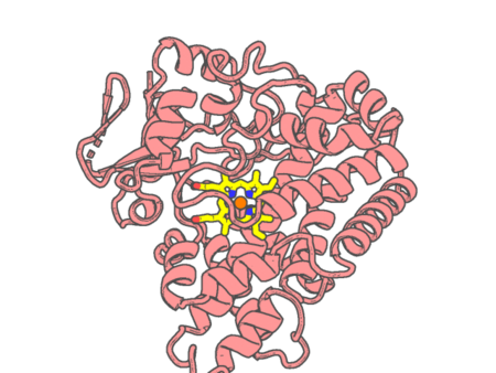 |
Experimental method | X-RAY DIFFRACTION |
|---|---|---|
| Structural data | available | |
| Non-standard amino acids | None | |
| Missing residues/ Structural gap | None / None | |
| Resolution | 2.2 Å | |
| Deposition date | 2016-02-22 |
| pdb_00005ibj | Crystal structure Mycobacterium tuberculosis CYP121 in complex with inhibitor fragment 6 | ||
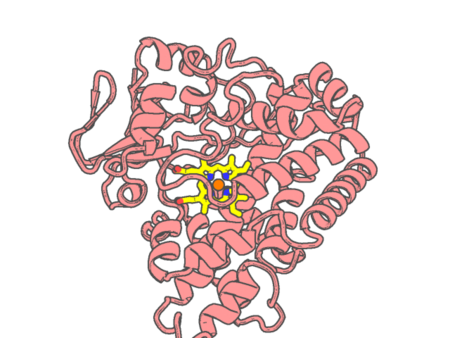 |
Experimental method | X-RAY DIFFRACTION |
|---|---|---|
| Structural data | available | |
| Non-standard amino acids | None | |
| Missing residues/ Structural gap | None / None | |
| Resolution | 2.5 Å | |
| Deposition date | 2016-02-22 |
| pdb_00005iki | CYP106A2 WITH SUBSTRATE ABIETIC ACID | ||
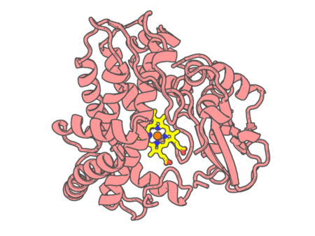 |
Experimental method | X-RAY DIFFRACTION |
|---|---|---|
| Structural data | available | |
| Non-standard amino acids | None | |
| Missing residues/ Structural gap | Exists / Exists | |
| Resolution | 2.4 Å | |
| Deposition date | 2016-03-03 |
| pdb_00005it1 | Streptomyces peucetius CYP105P2 complex with biphenyl compound | ||
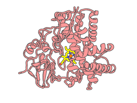 |
Experimental method | X-RAY DIFFRACTION |
|---|---|---|
| Structural data | available | |
| Non-standard amino acids | None | |
| Missing residues/ Structural gap | None / None | |
| Resolution | 2.1 Å | |
| Deposition date | 2016-03-16 |
| pdb_00005jhx | Crystal Structure of Fungal MagKatG2 at pH 3.0 | ||
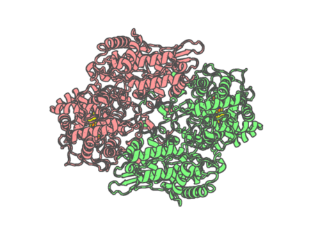 |
Experimental method | X-RAY DIFFRACTION |
|---|---|---|
| Structural data | available | |
| Non-standard amino acids | None | |
| Missing residues/ Structural gap | None / None | |
| Resolution | 1.4 Å | |
| Deposition date | 2016-04-21 |