PDB ID: pdb_00003n8y
- Structure of Aspirin Acetylated Cyclooxygenase-1 in Complex with Diclofenac
- Experimental method: X-RAY DIFFRACTION
- Resolution: 2.6 Å
- Deposition date: 2010-05-28
Number of assemblies: 1
| Structural data | modeled |
|---|---|
| Oligomeric count | 2 |
| label_asym_ids of protein | |
| label_asym_ids of heme | |
| label_asym_ids of the others |
C (NAG 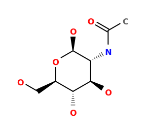 NAG
, BMA 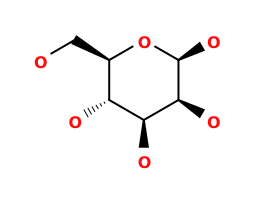 BMA
), D (NAGNAG
, NDG  NDG
, BMABMA
, MAN  MAN
), E (NAGNAG
, NDGNDG
), F (NAGNAG
, BMABMA
), G (NAGNAG
, NDGNDG
, BMABMA
), I (DIF  DIF
), J (BOG  BOG
), K (BOGBOG
), M (SAL  SAL
), N (DIFDIF
), O (BOGBOG
), P (BOGBOG
) |

Each chain information
| auth_asym_id | A |
|---|---|
| Residue indices (Uniprot) | 32-584 (32-584) |
| Mutations (UniProt numbering) | |
| Uniprot ID | P05979 →UniprotKB |
| S-S bonds | 36:47,37:159,41:57,59:69,569:575 |
| label_asym_ids of heme | H |
| Available structure | PDB |
| auth_asym_id | B |
|---|---|
| Residue indices (Uniprot) | 32-584 (32-584) |
| Mutations (UniProt numbering) | |
| Missing residues | 530SER-530SER |
| Nonstandard amino acids |
 OAS |
| Uniprot ID | P05979 →UniprotKB |
| S-S bonds | 36:47,37:159,41:57,59:69,569:575 |
| label_asym_ids of heme | L |
| Available structure | PDB and Modeled |
| RMSD of experimental residues after energy minimization | 0.10 Å |