
Number of data: 7660
Dataset: all data
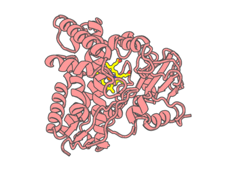
| pdb_00007qnn | Crystal structure of CYP125 from Mycobacterium tuberculosis in complex with inhibitor (surface entropy reduction mutant) | ||
 |
Experimental method | X-RAY DIFFRACTION |
|---|---|---|
| Structural data | available | |
| Non-standard amino acids | None | |
| Missing residues/ Structural gap | None / None | |
| Resolution | 2.47 Å | |
| Deposition date | 2021-12-21 |
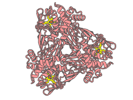
| pdb_00007qq2 | CRYSTAL STRUCTURE OF AS-ISOLATED S321M MUTANT OF THREE-DOMAIN HEME-CU NITRITE REDUCTASE FROM RALSTONIA PICKETTII | ||
 |
Experimental method | X-RAY DIFFRACTION |
|---|---|---|
| Structural data | available | |
| Non-standard amino acids | None | |
| Missing residues/ Structural gap | None / None | |
| Resolution | 2.1 Å | |
| Deposition date | 2022-01-06 |
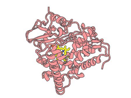
| pdb_00007qq7 | Crystal structure of CYP142 from Mycobacterium tuberculosis in complex with an inhibitor at partial occupancy with PEG | ||
 |
Experimental method | X-RAY DIFFRACTION |
|---|---|---|
| Structural data | available | |
| Non-standard amino acids | None | |
| Missing residues/ Structural gap | None / None | |
| Resolution | 1.6 Å | |
| Deposition date | 2022-01-06 |
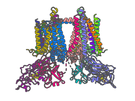
| pdb_00007qrm | Cryo-EM structure of catalytically active Spinacia oleracea cytochrome b6f in complex with endogenous plastoquinones at 2.7 A resolution | ||
 |
Experimental method | ELECTRON MICROSCOPY |
|---|---|---|
| Structural data | available | |
| Non-standard amino acids | None | |
| Missing residues/ Structural gap | Exists / Exists | |
| Resolution | None | |
| Deposition date | 2022-01-11 |
| pdb_00007qth | Crystal structure of the Shewanella oneidensis MR1 MtrC mutant C453A | ||
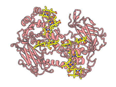 |
Experimental method | X-RAY DIFFRACTION |
|---|---|---|
| Structural data | available | |
| Non-standard amino acids | None | |
| Missing residues/ Structural gap | None / None | |
| Resolution | 1.9 Å | |
| Deposition date | 2022-01-14 |
| pdb_00007qu4 | Recombinant Human Fetal Hemoglobin mutant - alpha subunit mutations K11E,K56E,N78D,K90E | ||
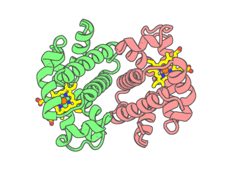 |
Experimental method | X-RAY DIFFRACTION |
|---|---|---|
| Structural data | available | |
| Non-standard amino acids | None | |
| Missing residues/ Structural gap | None / None | |
| Resolution | 1.66 Å | |
| Deposition date | 2022-01-17 |
| pdb_00007qwn | Crystal structure of CYP125 from Mycobacterium tuberculosis in complex with an inhibitor | ||
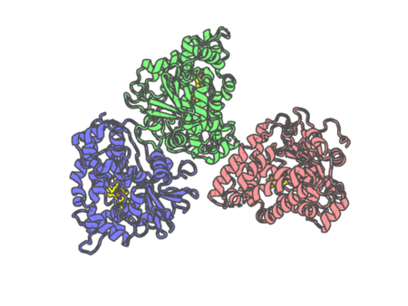 |
Experimental method | X-RAY DIFFRACTION |
|---|---|---|
| Structural data | available | |
| Non-standard amino acids | None | |
| Missing residues/ Structural gap | None / None | |
| Resolution | 1.93 Å | |
| Deposition date | 2022-01-25 |
| pdb_00007qyq | Crystal structure of a DyP-type peroxidase from Pseudomonas putida | ||
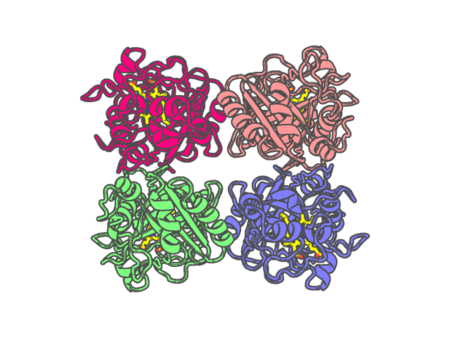 |
Experimental method | X-RAY DIFFRACTION |
|---|---|---|
| Structural data | available | |
| Non-standard amino acids | None | |
| Missing residues/ Structural gap | None / None | |
| Resolution | 2.599 Å | |
| Deposition date | 2022-01-28 |
| pdb_00007qyz | Crystal structure of a DyP-type peroxidase 6E10 variant from Pseudomonas putida | ||
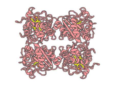 |
Experimental method | X-RAY DIFFRACTION |
|---|---|---|
| Structural data | available | |
| Non-standard amino acids | None | |
| Missing residues/ Structural gap | None / None | |
| Resolution | 2.452 Å | |
| Deposition date | 2022-01-30 |
| pdb_00007qza | Crystal structure of a DyP-type peroxidase 29E4 variant from Pseudomonas putida | ||
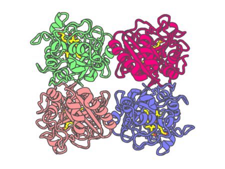 |
Experimental method | X-RAY DIFFRACTION |
|---|---|---|
| Structural data | available | |
| Non-standard amino acids | None | |
| Missing residues/ Structural gap | None / None | |
| Resolution | 2.698 Å | |
| Deposition date | 2022-01-30 |
| pdb_00007qze | SFX structure of dye-type peroxidase DtpB D152A variant in the ferryl state | ||
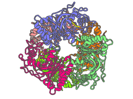 |
Experimental method | X-RAY DIFFRACTION |
|---|---|---|
| Structural data | available | |
| Non-standard amino acids | None | |
| Missing residues/ Structural gap | None / None | |
| Resolution | 1.9 Å | |
| Deposition date | 2022-01-31 |
| pdb_00007qzf | SFX structure of dye-type peroxidase DtpB D152A/N245A variant in the ferric state | ||
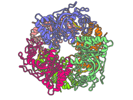 |
Experimental method | X-RAY DIFFRACTION |
|---|---|---|
| Structural data | available | |
| Non-standard amino acids | None | |
| Missing residues/ Structural gap | None / None | |
| Resolution | 2.2 Å | |
| Deposition date | 2022-01-31 |
| pdb_00007qzg | SFX structure of dye-type peroxidase DtpB N245A variant in the ferric state | ||
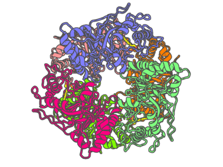 |
Experimental method | X-RAY DIFFRACTION |
|---|---|---|
| Structural data | available | |
| Non-standard amino acids | None | |
| Missing residues/ Structural gap | None / None | |
| Resolution | 2.1 Å | |
| Deposition date | 2022-01-31 |
| pdb_00007qzh | SFX structure of dye-type peroxidase DtpB D152A variant in the ferric state | ||
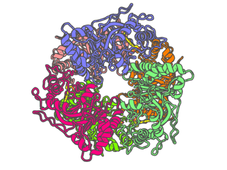 |
Experimental method | X-RAY DIFFRACTION |
|---|---|---|
| Structural data | available | |
| Non-standard amino acids | None | |
| Missing residues/ Structural gap | None / None | |
| Resolution | 1.92 Å | |
| Deposition date | 2022-01-31 |
| pdb_00007qzr | Structure of native leukocyte myeloperoxidase in complex with the Staphyloccal Peroxidase Inhibitor SPIN from Staphylococcus aureus | ||
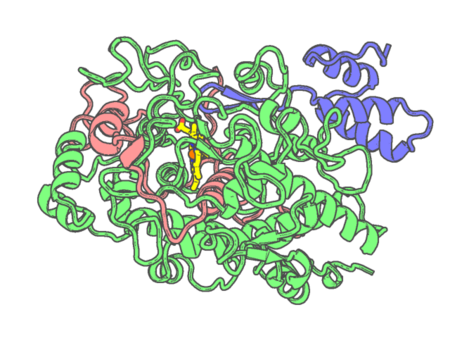 |
Experimental method | X-RAY DIFFRACTION |
|---|---|---|
| Structural data | available | |
| Non-standard amino acids | Included | |
| Missing residues/ Structural gap | None / None | |
| Resolution | 2.18 Å | |
| Deposition date | 2022-01-31 |
| pdb_00007r0w | 2.8 Angstrom cryo-EM structure of the dimeric cytochrome b6f-PetP complex from Synechocystis sp. PCC 6803 with natively bound lipids and plastoquinone molecules | ||
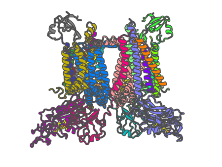 |
Experimental method | ELECTRON MICROSCOPY |
|---|---|---|
| Structural data | available | |
| Non-standard amino acids | None | |
| Missing residues/ Structural gap | Exists / Exists | |
| Resolution | None | |
| Deposition date | 2022-02-02 |
| pdb_00007r1i | Crystal structure of CYP125 from Mycobacterium tuberculosis in complex with an inhibitor | ||
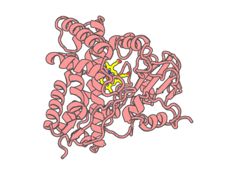 |
Experimental method | X-RAY DIFFRACTION |
|---|---|---|
| Structural data | available | |
| Non-standard amino acids | None | |
| Missing residues/ Structural gap | None / None | |
| Resolution | 2.24 Å | |
| Deposition date | 2022-02-03 |
| pdb_00007r2u | CRYSTAL STRUCTURE OF AS-ISOLATED Q262N MUTANT OF THREE-DOMAIN HEME-CU NITRITE REDUCTASE FROM RALSTONIA PICKETTII | ||
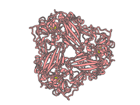 |
Experimental method | X-RAY DIFFRACTION |
|---|---|---|
| Structural data | available | |
| Non-standard amino acids | None | |
| Missing residues/ Structural gap | None / None | |
| Resolution | 1.5 Å | |
| Deposition date | 2022-02-05 |
| pdb_00007r3u | Crystal structure of CYP125 from Mycobacterium tuberculosis in complex with an inhibitor | ||
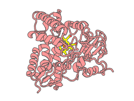 |
Experimental method | X-RAY DIFFRACTION |
|---|---|---|
| Structural data | available | |
| Non-standard amino acids | None | |
| Missing residues/ Structural gap | None / None | |
| Resolution | 1.86 Å | |
| Deposition date | 2022-02-07 |
| pdb_00007r3v | Crystal structure of bovine Cytochrome bc1 in complex with inhibitor CK-2-67. | ||
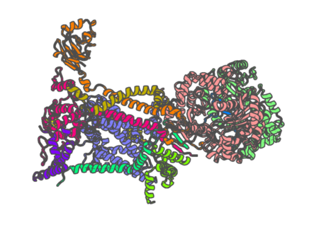 |
Experimental method | X-RAY DIFFRACTION |
|---|---|---|
| Structural data | available | |
| Non-standard amino acids | None | |
| Missing residues/ Structural gap | Exists / Exists | |
| Resolution | 3.2 Å | |
| Deposition date | 2022-02-07 |